Download
1 / 65
730 likes | 1.19k Views
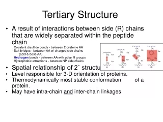
Chapter 6 Proteins: Secondary, Tertiary, and Quaternary Structure. Essential Question. How do the forces of chemical bonding determine the formation, stability , and myriad functions of proteins?. Outline. What noncovalent interactions stabilize protein structure?

E N D
Chapter 6Proteins: Secondary, Tertiary, and Quaternary Structure
Essential Question • How do the forces of chemical bonding determine the formation, stability, and myriad functions of proteins?
Outline • What noncovalent interactions stabilize protein structure? • What role does the amino acid sequence play in protein structure? • What are the elements of secondary structure in proteins, and how are they formed? • How do polypeptides fold into three-dimensional protein structures? • How do protein subunits interact at the quaternary level of protein structure?
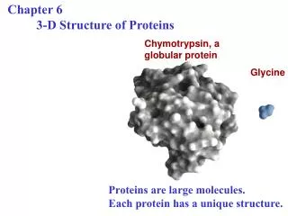
Protein Structure and Function Are Tightly Linked The three-dimensional structures of proteins and their biological functions are linked by several overarching principles: • Function depends on structure • Structure depends on sequence and on weak, noncovalent forces • The number of protein folding patterns is large but finite • Structures of globular proteins are marginally stable • Marginal stability facilitates motion • Motion enables function
6.1 What Noncovalent Interactions Stabilize the Higher Levels of Protein Structure? • Secondary, tertiary, and quaternary structure of proteins is formed and stabilized by weak forces • Hydrogen bonds are formed wherever possible • Hydrophobic interactions drive protein folding • Ionic interactions usually occur on the protein surface • van der Waals interactions are ubiquitous
Electrostatic Interactions in Proteins An electrostatic interaction between a positively charged lysine amino group and a negatively charged glutamate carboxyl group.
6.3 What Are the Elements of Secondary Structure in Proteins, and How Are They Formed? • The atoms of the peptide bond lie in a plane • All protein structure is based on the amide plane • The resonance stabilization energy of the planar structure is 88 kJ/mol • A twist about the C-N bond involves a twist energy of 88 kJ/mol times the square of the twist angle. • Rotation can occur about either of the bonds linking the alpha carbon to the other atoms of the peptide backbone
6.3 What Are the Elements of Secondary Structure in Proteins, and How Are They Formed? The amide or peptide bond planes are joined by the tetrahedral bonds of the α-carbon. The rotation parameters are φ and ψ. The conformations shown corresponds to φ= 180° and ψ= 180°.
Consequences of the Amide Plane Two degrees of freedom per residue for the peptide chain Angle about the Cα-N bond is denoted φ (phi) Angle about the Cα-C bond is denoted ψ (psi) The entire path of the peptide backbone is known if all φ and ψ angles are specified Some values of φ and ψ are more likely than others.
Some Values of φ and ψ Are Not Allowed Many of the possible conformations about an α-carbon between two peptide planes are forbidden because of steric crowding.
Steric Constraints on φ & ψ Unfavorable orbital overlap/steric crowding precludes some combinations of φ and ψ φ = 0°, ψ = 180° is unfavorable φ = 180°, ψ = 0° is unfavorable φ = 0°, ψ = 0° is unfavorable
Classes of Secondary Structure Secondary structures are local structures that are stabilized by hydrogen bonds • Alpha helices • Other helices • Beta sheet (composed of "beta strands") • Tight turns (aka beta turns or beta bends) • Beta bulge
The α-Helix Four different representations of the α-helix.
The α-Helix Numbers to Know • Residues per turn: 3.6 • Rise per residue: 1.5 Angstroms (0.15 nm) • Rise per turn (pitch): 3.6 1.5Å = 5.4 Angstroms • The backbone loop that is closed by any H-bond in an alpha helix contains 13 atoms • φ = −60 degrees, ψ = −45 degrees • The non-integral number of residues per turn was a surprise to crystallographers
The α-Helix in Proteins Two proteins that contain substantial amounts of α-helix.
Amino acids can be classified as helix-formers or helix breakers
The β-Pleated Sheet • The β-pleated sheet is composed of β-strands • Also first postulated by Pauling and Corey, 1951 • Strands in a β-sheet may be parallel or antiparallel • Rise per residue: • 3.47 Angstroms for antiparallel strands • 3.25 Angstroms for parallel strands • Each strand of a β-sheet may be pictured as a helix with two residues per turn
The β-Pleated Sheet A “pleated sheet” of paper with an antiparallel β-sheet drawn on it.
The β-Pleated Sheet H bonds in parallel and antiparallel β-sheets
Helix-Sheet Composites in Spider Silk Spider web silks are composites of α-helices and β-sheets. The radial strands of webs must be strong and rigid and have a higher percentage of β-sheets. The circumferential strands (termed capture silk) must be flexible and contain a higher percentage of α-helices.
The β-Turn (aka β-bend, or tight turn) • Allows the peptide chain to reverse direction • Carbonyl C of one residue is H-bonded to the amide proton of a residue three residues away • Proline and glycine are prevalent in β-turns • There are two principal forms of β-turns
The β-Turn The structures of two kinds of β-turns (also called tight turns or β-bends). Four residues are required to form a β-turn. Left: Type I; right: Type II.
6.4 How Do Polypeptides Fold into Three-Dimensional Protein Structures? Several important principles: • Secondary structures form wherever possible (due to formation of large numbers of H bonds) • Helices and sheets often pack close together • Peptide segments between secondary structures tend to be short and direct • Proteins fold so as to form the most stable structures. Stability arises from: • Formation of large numbers of intramolecular hydrogen bonds • Reduction in the surface area accessible to solvent that occurs upon folding
6.4 How Do Polypeptides Fold into Three-Dimensional Protein Structures? • Two factors lie at the heart of these principles: • Proteins are typically a mixture of hydrophilic and hydrophobic amino acids • The hydrophobic groups tend to cluster together in the folded interior of the protein
Fibrous Proteins • Much or most of the polypeptide chain is organized approximately parallel to a single axis • Fibrous proteins are often mechanically strong • Fibrous proteins are usually insoluble • Usually play a structural role in nature • Three types of fibrous protein are discussed here: • α-Keratin • β-Keratin • Collagen
α-Keratin • A fibrous protein found in hair, fingernails, claws, horns and beaks • Sequence consists of 311-314 residue alpha helical rod segments capped with non-helical N- and C-termini • Primary structure of helical rods consists of 7-residue repeats: (a-b-c-d-e-f-g)n, where a and d are nonpolar. • This structure promotes association of helices to form coiled coils
Collagen – A Triple Helix Principal component of connective tissue (tendons, cartilage, bones, teeth) • Basic unit is tropocollagen: • Three intertwined polypeptide chains (1000 residues each) • MW = 285,000 • 300 nm long, 1.4 nm diameter • Unique amino acid composition, including hydroxylysine and hydroxyproline • Hydroxyproline is formed by the vitamin C-dependent prolyl hydroxylase reaction.
Collagen – A Triple Helix The secrets of its a.a. composition... • Nearly one residue out of three is Gly • Proline content is unusually high • Unusual amino acids found: • 4-hydroxyproline • 3-hydroxyproline • 5-hydroxylysine • Pro and HyPro together make 30% of residues
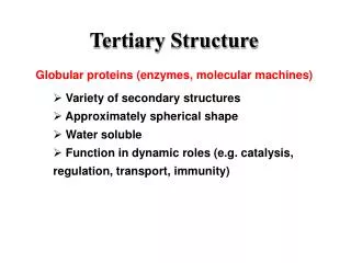
Globular Proteins Mediate Cellular Function • Globular proteins are more numerous than fibrous proteins • The diversity of protein structures in nature reflects the remarkable variety of functions they perform • Functional diversity derives in turn from: • The large number of folded structures that polypeptides can adopt • The varied chemistry of the side chains of the 20 common amino acids
Globular Proteins Some design principles • Helices and sheets make up the core of most globular proteins • Most polar residues face the outside of the protein and interact with solvent • Most hydrophobic residues face the interior of the protein and interact with each other • Packing of residues is close • However, ratio of van der Waals volume to total volume is only 0.72 to 0.77, so empty space exists • The empty space is in the form of small cavities
“Random coils” are not random • The segments of a protein that are not helices or sheets are traditionally referred to as “random coil”, although this term is misleading: • Most of these segments are neither coiled or random • They are usually organized and stable, but don’t conform to any frequently recurring pattern • Random coil segments are strongly influenced by side-chain interactions with the rest of the protein
Globular Proteins The structure of ribonuclease, showing elements of helix, sheet and random coil.
Protein surfaces are complex The surfaces of proteins are complementary to the molecules they bind.
Waters on the Protein Surface Stabilize the Structure The surfaces of proteins are ideally suited to form multiple H bonds with water molecules.
α-Helices May be Polar, Nonpolar or Amphiphilic The so-called helical wheel presentation can reveal the polar or nonpolar character of α-helices.
Protein domains are nature’s modular strategy for protein design • Proteins composed of about 250 amino acids or less often have a simple, compact globular shape • Larger globular proteins are typically made up of two or more recognizable and distinct structures, termed domains or modules – compact, folded protein structures that are usually stable by themselves in aqueous solution • Domains may consist of a single continuous portion of the protein sequence (see Figure 6.23) • In some proteins, the domain sequence is interrupted by a sequence belonging to another part of the protein (Figure 6.24)
Many proteins are composed of several distinct domains Several protein modules used in the construction of complex multimodule proteins.
Classification Schemes for the Protein Universe Are Based on Domains • Common features of SCOP and CATH: • Class is determined from overall composition of secondary structure elements in a domain • Fold describes the number, arrangement, and connections of these secondary structure elements • Superfamily includes domains of similar folds and usually similar functions • Family usually includes domains with closely related amino acid sequences
Structure and Function are Not Always Linked • Because structure depends on sequence, and because function depends on structure, it is tempting to imagine that all proteins of similar structure should share a common function, but this is not always true • Some proteins of similar domain structure have different functions • Some proteins of similar function possess very different structures
Denaturation Leads to Loss of Protein Structure and Function • The cellular environment is suited to maintaining the weak forces that preserve protein structure and function • External stresses – heat, chemical treatment, etc. – can disrupt these forces in a process termed denaturation – the loss of structure and function • The cooking of an egg is an everyday example • Ovalbumin, the principal protein in egg white, remains in its native structure up to a characteristic melting temperature, Tm • Above this temperature, the structure unfolds and function is lost
Denaturation Leads to Loss of Protein Structure and Function The proteins of egg white are denatured during cooking. More than half of the protein in egg white is ovalbumin.
Denaturation Leads to Loss of Protein Structure and Function Proteins can be denatured by heat, with commensurate loss of function.
Denaturation Leads to Loss of Protein Structure and Function Proteins can be denatured (unfolded) by high concentrations of guanidine-HCl or urea. The denaturation of chymotrypsin is plotted here.
Anfinsen’s Classic Experiment Proved that Sequence Determines Structure Ribonuclease can be unfolded by treatment with urea. β-Mercaptoethanol (MCE) cleaves disulfide bonds. Anfinsen showed that ribonuclease structure (and function) could be restored under appropriate conditions.
Is There a Single Mechanism for Protein Folding? • How a protein achieves its stable, folded state is a complex question • Levinthal’s paradox demonstrates that proteins cannot fold by sampling all possible conformations • This implies that proteins actually fold via specific “folding pathways” • What factors play a role in protein folding processes?
Postulated Themes of Protein Folding • Secondary structures – helices, sheets, and turns – probably form first • Nonpolar residues may aggregate or coalesce in a process termed a hydrophobic collapse • Subsequent steps probably involve formation of long-range interactions between secondary structures or involving other hydrophobic interactions • The folding process may involve one or more intermediate states, including transition states and what have become known as molten globules
The Protein Folding Energy Landscape Ken Dill has suggested that the folding process can be pictured as a funnel of free energies. The rim at the top represents the many unfolded states. Polypeptides ‘fall down the wall of the funnel’ to ever fewer possibilities and lower energies as they fold.
Motion is Important for Globular Proteins • Protein are dynamic structures – they oscillate and fluctuate continuously about their average or equilibrium structures • This flexibility is essential for protein functions, including: • Ligand binding • Enzyme catalysis • Enzyme regulation