
BACKGROUND
10 weeks. 16 weeks. 24 weeks. 30 weeks. 10 weeks. 16 weeks. 24 weeks. 30 weeks. Longitudinal study of cartilage and subchondral bone changes in a spontaneous animal model of knee OA.

BACKGROUND
E N D
Presentation Transcript
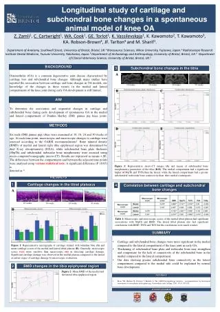
10 weeks 16 weeks 24 weeks 30 weeks 10 weeks 16 weeks 24 weeks 30 weeks Longitudinal study of cartilage and subchondral bone changes in a spontaneous animal model of knee OA Z. Zamli1, C. Cartwright1, WA. Cook1, GE. Torlot1, K. Vassilevskaja1, K. Kawamoto2, T. Kawamoto3, KA. Robson-Brown4, JF. Tarlton5 and M. Sharif1. Department of Anatomy, Southwell Street, University of Bristol, Bristol, UK.1 Bioresource Sciences, Nihon University, Fujisawa, Japan.2 Radioisotope Research Institute Dental Medicine, Tsurumi University, Yokohama, Japan.3 Department of Archaeology and Anthropology, University of Bristol, Bristol, UK.4 Department of Clinical Veterinary Science, University of Bristol, Bristol, UK.5 BACKGROUND BACKGROUND BACKGROUND BACKGROUND BACKGROUND BACKGROUND 3 Subchondral bone changes in the tibia Osteoarthritis (OA) is a common degenerative joint disease characterized by cartilage loss and subchondral bone changes. Although many studies have reported the association between cartilage and bone changes in OA models, our knowledge of the changes in these tissues in the medial and lateral compartments of the knee joint during early OA development is still limited. A B C AIM AIM AIM AIM AIM To determine the association and sequential changes in cartilage and subchondral bone during early development of spontaneous OA in the medial and lateral compartment of Dunkin Hartley (DH) guinea pig knee joints. METHODS D E Six male (DH) guinea pigs tibias were examined at 10, 16, 24 and 30 weeks of age. At each time point, macroscopic and microscopic changes to cartilage were assessed according to the OARSI recommendations1. Bone mineral density (BMD) of medial and lateral right tibia epiphyseal region was determined by dual X-ray absorptiometry (DXA), while subchondral bone plate thickness (SbpTh) and subchondraltrabecular bone morphometry were assessed using micro-computed tomography (micro-CT). Results are expressed as means ± SE. The differences between the compartments and between the adjacent time points were analysed using various statistical tests. A significant difference (P≤0.05) is denoted as *. Figure 3: Representative micro-CT images (A) and means of subchondral bone morphometry parameters of the tibia (B-E). The medial compartment had a significant higher ofSbpTh and TbTh than the lateral, while the lateral compartment had a greater subchondraltrabecularbone connectivity than their medial counterparts. RESULTS 1 Cartilage changes in the tibial plateaus 4 Correlation between cartilage and subchondral bone changes A B Table 1: Macroscopic and microscopic scores of the medial tibial plateau had significant associations with SbpTh and BMD. The lateral tibial plateau also had significant correlations with BMD, TbTh and TbN but the correlations were much weaker. SUMMARY • Cartilage and subchondral bone changes were more significant in the medial compared to the lateral compartment of the knee joint in early OA. • Thickening of subchondral bone plate and trabecular bone may strengthen and compensate for the lack of connectivity of the subchondral bone in the medial compared to the lateral compartment. • Our data showing greater subchondral bone connectivity in the lateral compartment compared to the medial side could be explained by normal bone development. Figure 1: Representative micrographs of cartilage stained with toluidine blue (A) and mean cartilage scores of the medial and lateral tibial plateau (B). Generally, microscopic scores were more sensitive than macroscopic one in detecting cartilage damage. Significant cartilage damage was observed in the medial plateau compared to the lateral at earlier stages of cartilage damage by microscopic evaluation. 2 BMD changes in the tibia epiphyseal region Figure 2: Mean BMD of the medial and the lateral tibia epiphyseal region. REFERENCE • Kraus VB, Huebner JL, DeGroot J, Bendele A. The OARSI histopathology initiative - recommendations for histological assessments of osteoarthritis in the guinea pig. Osteoarthritis and Cartilage. 2010; 18 (3): S35-S52. ACKNOWLEDGEMENT


