Download
1 / 20
280 likes | 736 Views
Movement Disorders: Basal Ganglia. Lecture Outline Identification and localization of major basal gangliar components. Functional relations among the major basal gangliar components. Synaptic organization contributing to direct and indirect motor loops.

E N D
Lecture Outline • Identification and localization of major basal gangliar components. • Functional relations among the major basal gangliar components. • Synaptic organization contributing to direct and indirect motor loops. • Implications of activity within components of the direct and indirect motor loops for cortical activity and hence movement. • The nature and causes of hypokinesia. • The nature and causes of hyperkinesia. • Therapies for Parkinson disease. • Therapies for Huntington disease. 2
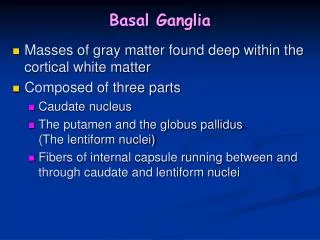
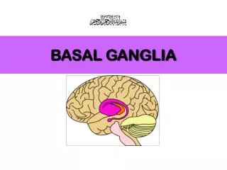
Identification of Major Basal Nuclear Components • The caudate and putamen are continuous with each other (forming the striatum). • The globuspallidus has a External (lateral) part (GPe), internal (medial) part GPi) • The putamen and globuspallidus form the lentiform nucleus. • The claustrum and amygdala (not shown) are also often considered as basal ganglia. • The subthalamus and thalamus participate in basal gangliar function.
Structural Relations Among Basal Ganglia • The caudate follows the lateral ventricle, with its tail terminating at the amygdala. • The lentiform nucleus sits lateral to the internal capsule.
Principles of Basal Gangliar Influence on Motor Activity •Basal ganglia receive inputs from cerebral cortex. •Basal ganglia modulating the thalamus. •Thalamic output regulates motor cortex. •Through indirect influences on motor cortex, basal ganglia participate in the initiation and control of voluntary movement.
Vital Modulatory Input • The substantianigra projects to the striatum to mediate the release of dopamine. • Dopamine regulates basal nuclear function, thus participating indirectly in regulation of cortical control of movement.
Pathways in the basal ganglia Almost all areas of the cerebral cortex project topographically onto the striatum, including a critical input from the motor cortex. The striatum then communicates with the thalamus and then back to the cortex via two different pathways.
Direct Basal Gangliar Loop • Striatal output suppresses activity in the GPi. • Decreased output from the GPi increases thalamic activity. • Increased thalamic activity increases cortical activity to facilitate movement. (note D1 receptors on striatal cells receiving dopaminergic projections from the substantianigra [SN]).
Indirect Basal Gangliar Loop • Striatal output suppresses activity in the GPe. • Decreased output from the GPe increases output from the Sth. • Increased output from the Sth increases activity within the GPi. • Increased activity within the GPi inhibits the thalamus. • Decreased thalamic output decreases cortical activity, thereby diminishing movement. (note D2 receptors on striatal cells receiving dopaminergic projections from the substantianigra [SN]).
Balanced Activity in Direct andIndirect Basal Gangliar Loops • Optimized communication between upper and lower motor neurons depends on delicately balanced activities of direct and indirect basal gangliar circuits. • Imbalances in activity of direct and indirect circuits yield either hypo- or hyperkinesis.
NigrostriatalDopaminergic Insufficiencyin Parkinson Disease • Insufficient release of dopamine alters activity in both direct and indirect pathways so as to decrease inhibition of the globuspallidus internal. • The net effect is increased inhibition of the thalamus, leading to decreased cortical activity and hypokinesis.
Other Parkinsonian Conditions • Drug-induced • Antipsychotic drugs • Depletors of DA stores (e.g., reserpine) • Toxic contaminants (MPTP) • Vascular • Strokes disrupting nigrostriatal pathway. • Traumatic • Multiple blows to the head can damage the midbrain, yielding a syndrome with parkinsonian features. • Postencephalitic • Viral encephalitis leads to nigral degeneration. • Neoplastic • Tumors can disturb the nigrostriatal system.
Striatal Neuronal Degeneration and Huntington Disease • Destruction of striatal neurons expressing D2 receptors. • Reduced inhibition of thalamus via indirect pathway. • Increased cortical activity, leading to hyperkinesis. (note faint “X” indicating loss of striatal neurons expressing D2 receptors).
Hemiballismus • Flinging and rotational movements of the limbs arise from contralateral subthalamic injury. • Reduced excitation of the globus pallidus internal disinhibits the thalamus and hece the cortex. (note faint “X” indicating damage to the subthalamus (Sth).
Movement Disorders: Cerebellum
Cerebellar Functions • Synergy of movement, whereby many elemental • muscular contractions are collectively coordinated • to create purposeful (perhaps highly complex) • movement (e.g., walking). • Posture, whereby the body is oriented • appropriately relative to the vector of gravity. • Muscular tone, whereby muscles • exhibit appropriate basal levels of • contraction and thus determine posture.
Cerebellar Function and Dysfunction: Basic Rules • Cerebellum regulates ipsilateral body. • Acute cerebellar damage oftens yields • pronounced motor deficits (ipsilaterally).
Lobar Structure • Anterior lobe: • Posterior lobe • Flocculonodular lobe Cerebellar hemisphere indirectly regulates ipsilateral body. Multiple motor homunculi present. Midline regulates trunk. More lateral structures regulate limbs.
Three Functional Zones • Vestibulocerebellum for • regulation of balance and • eye movements • (linked to CN VIII and FN • lobe). • Spinocerebellum regulates • trunk and limbs (vermis and • paravermis). • pontocerebellumregulates • learning, planning, precise • timing (lateral hemispheres).