Overview of Blood, Nerve, Muscle Tissues, and Gland Functions
320 likes | 441 Views
This comprehensive guide explores the various types of tissues in the body, focusing on blood, nerve, and muscle tissues, along with endocrine and exocrine gland functions. Blood serves critical roles in transporting oxygen, immune response, and clotting. Nerve tissue facilitates rapid signal transmission, while muscle tissue is responsible for movement and generating body heat. Understanding these tissues' structures and functions is essential for grasping human biology and its complexities.

Overview of Blood, Nerve, Muscle Tissues, and Gland Functions
E N D
Presentation Transcript
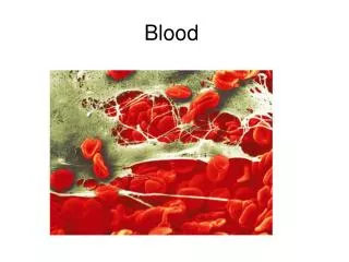
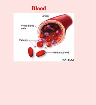
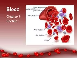
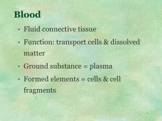
Blood • Fluid connective tissue • Function: transport cells & dissolved matter • Ground substance = plasma • Formed elements = cells & cell fragments
1-RBC 2-WBCs 4-Platelets
Blood • Erythrocytes (RBCs) – O2 & CO2 • Leukocytes (WBCs) – immune functions • Neutrophils • Eosinophils, • Basophils • Lymphocytes • Monocytes • Platelets – cell fragments, clotting & vessel growth factor
Nerve Tissue • Excitable tissue – has membrane potential • Voltage difference between outer & inner membrane • Results in rapid transition of signals to other cells • Located brain, spinal cord, nerves, ganglia
Nerve Tissue • Neuron (nerve cell) • Soma (cell body) • Dendrites (input) • Axon (output) • Neuroglia or Glial cells (white matter)
Muscle Tissue (Table 5.10) • Specialized contractile cells (spindle) • Exert physical force on other tissues • Important source of body heat (contraction) • 3 types of muscle: • Skeletal • Cardiac • Smooth
Skeletal Muscle • Long cylindrical cells – muscle fibers • Striated (alternating light & dark bands) • Voluntary (conscious control over contraction)
Cardiac Muscle • Only in the heart • Shorter cells – myocytes • Joined together at ends by intercalated discs • Striated & involuntary
Smooth Muscle • Short fusiform cells • Non-striated with only one central nucleus • Involuntary contraction • Primary muscles of viscera
Intercellular Junctions • Connections between cells • Means of Communication • Resists stress to tissue
Tight Junctions • Encircles cell near Apex • Zipperlike with grooves and ridges • Prevents passage between cells
Desmosomes • “Snap-like” • Doesn’t encircle cell • Located in epidermis, cardiac myo, & cervix
Gap Junctions • Protein ring around water-filled pore • small solutes pass through cell to cell • Intercalated discs & smooth myo • Allows ions flow for electrical excitation
Endocrine & Exocrine Glands • Exocrine glands secrete through duct • Sweat, mammary, tears • Endocrine glands secrete hormones directly into bloodstream • Pituitary, thyroid, adrenals • Some organs have both kinds • liver, gonads, pancreas
Exocrine Gland Structure • Enclosed in fibrous capsule • Septa or trabeculae divide glands into lobes • Stroma is framework (CT) • Parenchyma are active cells
Types of Exocrine Glands • Simple unbranched duct • Compound branched duct • Shape of gland: acinar - secretory cells form dilated sac tubuloacinar - both tube and sacs
Types of Secretions • Serous glands • thin, watery secretions • sweat, milk, tears and digestive juices • Mucous glands • Mucin + water = mucus • Mixed glands contain both types • Some salivary glands • Cytogenic glands release whole cells • sperm & egg cells
Methods of Exocrine Secretion • Merocrine (eccrine) Glands • Secretes via exocytosis • Tears, pancreas, gastric, sweat • Apocrine (also merocrine) • Holocrine • Entire cell disintegrates • Sebaceous glands of scalp
Mucous Membranes Lamina propria = areolar connective tissue
Tissue Growth • Hyperplasia = growth by cell multiplication • Hypertrophy = growth by increasing size • muscle grow through exercise • Neoplasia = tumor growth (benign or malignant) abnormal, nonfunctional tissue
Changes in Tissue Types • Differentiation • Development of unspecialized tissues into specialized mature types • mesenchyme becomes muscle • Metaplasia • Mature tissue change into another • Normal: simple cuboidal tissue before puberty changes to stratified squamous after puberty • Abnormal: smokers – pseudostratified columnar of bronchi into stratified squamous
Stem Cells-undifferentiated cells • Embryonic stem cells • Totipotent (any cell type possible) • source = cells of very early embryo • Pluripotent (tissue types only possible) • source = cells of inner cell mass of embryo • Adult stem cells • Multipotent (several possible cell type – but not all) • Unipotent (only epidermal cells produced)
Tissue Repair • Regeneration • replacement of damaged cells with functioning cells • skin injuries and liver regenerate • Fibrosis • replacement of damaged cells with scar tissue • Loss of function - healing muscle injuries, scarring of lung tissue in TB or healing of severe cuts and burns of the skin • Keloid - excessive fibrosis during healing (raised shiny scars)
Clot & Scab forms on surface Macrophages start to clean up debris Antibodies, clotting factors and WBCs drawn wound
New capillaries – Fibroblasts deposit collagen – May last 2 weeks Epithelials multiply beneath scab Epi-layer thickens Connective tissue scars (fibrosis) Phase may last 2 years
Tissue Shrinkage and Death • Atrophy = loss of cell size or number • disuse atrophy from lack of use (leg in a cast) • Necrosis = pathological death of tissue • gangrene – tissue death from lack of blood • infarction - death of heart tissue from lack of blood • Apoptosis = programmed cell death • cells shrink and are phagocytized (no inflammation)