Download

1 / 39
680 likes | 2.21k Views
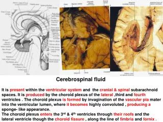
2. Introduction. Cerebrospinal fluid rhinorrhea/otorrheaAbnormal communication between the subarachnoid space and nose/temporal boneComplications highMeningitis/brain abscessChallenge for diagnosis and treatmentImportant for otolaryngologists. 3. CSF Rhinorrhea. Connection of SA space to nos

E N D
1. 1 Cerebrospinal Fluid Rhinorrhea and Otorrhea Russell D. Briggs, M.D.
Matthew Ryan, M.D.
2. 2 Introduction Cerebrospinal fluid rhinorrhea/otorrhea
Abnormal communication between the subarachnoid space and nose/temporal bone
Complications high
Meningitis/brain abscess
Challenge for diagnosis and treatment
Important for otolaryngologists
3. 3 CSF Rhinorrhea Connection of SA space to nose/sinuses
Diverse etiologies
Iatrogenic� ESS
Blunt trauma� CHI or skull fractures
Increased intraventricular pressure
Tumors, post infectious/traumatic hydrocephalus
Arachnoid granulations
4. 4 CSF Rhinorrhea History and PE
Unilateral watery rhinorrhea
Increases with valsalva and posture
May see leak/encephalocele with endoscope
Collect fluid
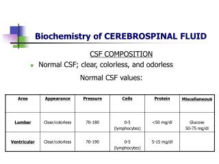
5. 5 CSF Rhinorrhea Ensure it a CSF leak
Testing of secretions
Beta-2-transferrin � highly specific
Glucose/protein determination
Electronic nose
6. 6 CSF Rhinorrhea Most important step� identify the site
High resolution CT of sinuses (1mm)
Coronal good for anterior skull base
Axial good for posterior wall frontal sinus
Problem is volume averaging
Look in cribiform niche and lateral wall of sphenoid sinus
7. 7 High resolution CT
8. 8 High Resolution CT
9. 9 CT Cisternogram Optimal imaging technique
False negative if no active leak
Obtain if HRCT fails to show the defect
10. 10 Magnetic Resonance Imaging MR cisternography�misnomer as no intrathecal contrast
Poor bony detail
Uses highly T2 weighted images
New method with intrathecal gad
Encephaloceles
11. 11 Radioisotope cisternography Many false positives and negatives
Fallen out of favor
No anatomic detail
For selected cases when leak not identified
Cottonoids in MM, SE recess
Removed in 24 hours and tested
If positive� intrathecal florescein
12. 12 Intrathecal Florescein IF leak not identified and strong suspicion
Combined with endoscopic surgical approach
Complications
Topical use
13. 13 Treatment of CSF Rhinorrhea Most resolve (after trauma/surgery)
Bed rest, head elevation, stool softeners
Possible lumbar drain/spinal taps
Prophylactic antibiotics
Surgical repair
Extensive intracranial injury
Intraoperative identification
Do not respond to conservative measures
14. 14 Surgical Treatment Intracranial
Time tested
Allows direct visualization
Well vascularized flaps
Success about 75%
High morbidity (anosmia, edema, hemorrhage, incision, hospital stay)
15. 15 Surgical Treatment Extracranial
Uses facial incisions for direct visualization
Success about 80%
Morbidity� facial scarring
16. 16 Surgical Treatment Endoscopic intranasal
Preferred method of repair
Successful 83-94% (average 90%)
Different techniques used
Overlay vs. Underlay techniques
Composite grafts
Dependent on size and location of defect
Sphenoid sinus
17. 17 Surgical Techniques
18. 18 Surgical Techniques
19. 19 Surgical Techniques Use gelfoam and gelfilm (>90%)
Use nasal packing (100%)
Consider fibrin glue (>50%)
Consider lumbar drain
3-5 days
Not required
BR, stool softeners, antibiotics
20. 20 CSF Otorrhea Connection of SA space and TB
Acquired etiology is most common
Trauma (temporal bone fracture), post-operative, infections, neoplasms
Congenital etiologies
Mondini deformities, wide CA, patent Hyrtel�s fissure, wide fallopian canal
Arachnoid granulations (�Spontaneous�)
21. 21 Temporal Bone Fractures Most common cause of CSF otorrhea
Longitudinal vs. Transverse
CSF from ear or nasopharynx
HRCT
Send fluid for beta-2-transferrin
Bed rest, head elevation, stool softeners, occ lumbar drain, sterile cotton, antibiotics (no drops)
22. 22 Temporal bone fractures Brodie and Thompson (1997)
Review of 820 TB fractures
122 with CSF leak
95 closed in first week, 21 in second week, only 5 drained over two weeks
Seven patients had surgery
Check scan and audiogram
9 developed meningitis
?Abx
23. 23 Spontaneous CSF Otorrhea May be subtle
Two types
Preformed bony pathway� present early
Meningitis after AOM
Resistant MEE� recognized after MT
Congenital defect (arachnoid granulations)
Villi enlarge, weight of temporal lobe
Bone erosion� present over age 50
MEE
24. 24 Spontaneous CSF Otorrhea
25. 25 Spontaneous CSF Otorrhea
26. 26 Spontaneous CSF Otorrhea
27. 27 Spontaneous CSF Otorrhea Beta-2-transferrin
HRCT
CT cisternogram
MR cisternogram
Surgical repair
28. 28 Surgical Techniques Middle fossa defects
Middle fossa craniotomy with extradural elevation� avoids ossicular problems
Transmastoid
Posterior fossa defects
Transmastoid/fat obliteration of mastoid
Others
29. 29 Conclusions Get a good history and PE
Test the fluid (if possible)
Find the site of the the leak
Radiographically
Treat it surgically if necessary
30. 30 Case Report 45 yobf presents with �headache and my neck hurts�
31. 31 Case Report 45 yobf presents with �headache and my neck hurts�
Worsening for 2 weeks
Photophobia, N/V
32. 32 Case Report 45 yobf presents with �headache and my neck hurts�
Worsening for 2 weeks
Photophobia, N/V
PMH: meningitis 6 months prior, AR
PSH: hysterectomy
Meds: Flonase� not helping� constant drainage
SH/FH/ROS: NC
33. 33 Case Report Physical Exam
Positive Kernig�s and Brudinski�s
Some clear rhinorrhea and hypertrophied turbs bilaterally
Sits forward and clear fluid from right nare
Otherwise normal H/N exam
34. 34 Case Report Labs: WBC= 20.2 with left shift, remainder essentially OK
35. 35 Case Report Consult to neurology made
LP� cloudy fluid,many PMN�s
Streptococcus pneumoniae
Placed on appropriate abx
Improving
36. 36 Case Report
37. 37 Case Report
38. 38 Case Report
39. 39 Case Report Did not respond to conservative measures
Taken to surgery
Endoscopically identified leak (3-4mm)
Three layer repair
Lumbar drain in for 7 days
Packing in for 7 days

![CEREBRAL CIRCULATION AND CEREBROSPINAL FLUID [CSF]](https://cdn2.slideserve.com/4005143/slide1-dt.jpg)