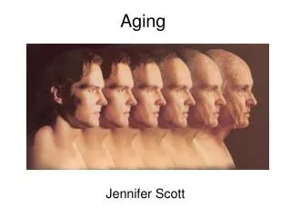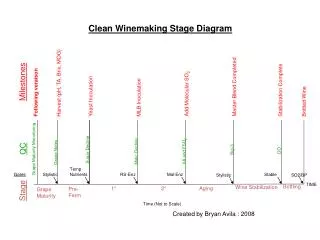
Aging
Aging. Musculoskeletal Function and Aging. Factors that Accentuate Aging. Lack of exercise prolonged immobilizations from injuries Medications : glucocorticoids, thyroxine, etc. Underlying disease states :

Aging
E N D
Presentation Transcript
Factors that Accentuate Aging • Lack of exercise • prolonged immobilizations from injuries • Medications: glucocorticoids, thyroxine, etc. • Underlying disease states: • Metabolic dysfunction: hyperthyroidism, hyperparathyroidism, hypercalcinuria, paraneoplastic neoplasias etc. • Neuropathies • Arthritis, other rheumatological disorders • Gastrectomy, other gastrointestinal disorders • Risk factors: • Alcoholism • Cigarette smoking
Bone Mass and Remodelling Replacement of old bone by new • The skeleton undergoes periodic remodeling • Responsible for its complete regeneration every 10 years • Remodeling primarily by a team of osteoclasts and osteoblasts that together comprise the basic multicellular unit (BMU) • At the leading edge of the BMU, osteoclasts adhere to bone and subsequently remove it by acidification and proteolytic digestion. Osteoclasts then leave the resorption site, and osteoblasts move in and secrete osteoid, which is eventually mineralized into new bone • Bone mass is preserved through a remarkably tight balance between resorption and formation. • In aging, the osteoclast activity is greater than osteoblast activity and results in net bone loss
Bone Loss: Androgen Effect • From about age 45, bone density (bone mass per unit volume) progressively decreases in both sexes, but more rapidly in women • A decline in sex hormones and aging itself both contribute to the loss of bone density • Testosterone and estrogen regulate some development and death (apoptosis) of osteoclasts and osteoblasts • Altered production of cytokines and altered responsiveness of bone marrow cell progenitors to cytokines. • For example, the production of interleukin-6 (IL-6) by osteoblasts is inhibited by estrogen and androgen.
Bone Loss: Men vs. Women • In men: testosterone production declines gradually, so bone loss is linear and slow • In women: • a rapid phase of bone loss occurs during the first 5 to 10 years after menopause • women, during their growing years and particularly during puberty, accumulate less skeletal mass than men, resulting in smaller, narrower, more fragile bones with thinner cortices • In old age, bone loss is greater among women than men, and the incidence of bone fractures is X2-3 higher
Bone Loss: Menopause vs. Aging Post-menopausal changes in bone loss eventually overlap with the effects of aging in women and are hard to differentiate
Bone Loss: Aging • Axial skeleton (cancellous [trabecular]) • Appendicular skeleton(cortical) (A) Autopsy specimen from young bone (B) Specimen from old bone showing marked reduction of cancellous bone Schematic depiction of bone reduction in aging cancellous bone: Normal bone undergoes resorption (shaded area), longitudinal trabeculae become thinner and some transverse trabeculae disappear (bottom)
Bone Loss: Aging Changes in cortical bone: reduced thickness and increased porosity. Women have thinner cortices than men do, so the effect of cortical thinning is more pronounced in women. Relationship between bone mass, age and gender Top of figure: Cross-section of the diaphysis of the femur with bone mass configuration shown
Bone Aging Summary • Amount of bone formed during remodeling decreases with age in both sexes • Consistent decrease in wall thickness, especially in trabecular bone • Formation of osteoblasts decreases • Rate of bone formation decreases • Bone mineral density decreases • Formation of adipocytes in the bone marrow increases • Changes result in the osteopenia • Vertebral and hip fractures are typical of senescent bone loss
Bone: Biomechanics of Aging Quantifying changes of aging is difficult and there is a lot of variation between different studies
Bone: Biomechanics of Aging Adult young and old human tibia tested in tension • Note the much earlier point of failure of the older bone and the lower curve for bone from women • Change is a factor of: • Increased brittleness • Diminished ability to deform
Cancellous Bone: Biomechanics of Aging Decrease in failure stress correlates with degree of bone thinning and loss of trabeculae
Muscle Loss • Between ages 30 and 75, lean body mass decreases, primarily due to loss of skeletal muscle mass • In healthy young persons, 30% of body weight is muscle, 20% is adipose tissue, and 10% is bone • By age 75, about 15% of body weight is muscle, 40% is adipose tissue, and 8% is bone • The number and size of muscle fibers progressively decrease. This process is called sarcopenia • The age-related loss of muscle fibers correlates with a loss of maximum isometric contraction force, which decreases 20% by the 6th decade and 50% by the 8th decade.
Type II Muscle Loss • Type II fibers participate in sudden powerful muscle contractions, whereas type I fibers function to maintain posture and to perform rhythmic, endurance-type exercises • The faster-contracting type II muscle fibers decrease to a greater extent than do the slower-contracting type I muscle fibers • Despite age-related reductions in muscle strength, muscle functional ability is similar in older and younger adults - usually, healthy elderly persons can easily climb stairs, rise from a squatting position, walk along a straight line, hop on either foot, and perform typical activities of daily living.
Muscle Loss: Age-Related Factors • Reduced levels of physical activity, decreasedroutine performance of vigorous muscular work, increased rates of immobilization or deconditioning from injuries or disease • Changes in the central or peripheral nervous system, possibly beginning during middle age, that may lead to a loss of motor units • Reduced rate of skeletal muscle protein synthesis • Relative deficiency of anabolic hormones, such as growth hormone, insulin-like growth factor I (IGF-I), testosterone, and dehydroepiandrosterone (DHEA) • Greater dietary protein requirements coupled with reduced protein intake
Articular Cartilage: Aging • It is difficult to differentiate between the changes of senescence and the changes of degenerative processes • Some degeneration of the knee joint is observable in every subject beyond the age of 15 • However, changes seen in thirty-year olds may not be seen in eighty-year olds so it is difficult to attribute changes to aging • The majority of changes are observed in weight-bearing portions of articular cartilage that are not covered by the menisci
Articular Cartilage: Aging • In some cases, knee cartilage thins rapidly or remains morphologically normal • There is no evidence that thickness of articular cartilage changes with age • Most common changes seen are: non-progressive fibrillation, change in color (yellow-brown tinge), thickness or biomechanical properties • In most cases, articular cartilage appears visually intact but demonstrates significant biomechanical changes
Cartilage: Biomechanics of Aging • The number of collagen fibers stays the same but may become thinner and finer resulting in altered mechanics • There is a decrease in: • Static tensile fracture strength • Static stiffness • Tensile fatigue strength
Ligaments & Tendons: Aging • Proliferative capacity and synthetic activity of fibroblasts decreases with age • These changes partially explain diminished healing capacity with age • Fibroblasts are responsible for the maintenance of connective tissues like ligaments and tendons • The demonstrable decrease in the strength of ligaments and tendons increases the risk that these structures will fail with age.
Ligaments: Biomechanics of Aging • Diminshed fibroblast activity directly results in inadequate maintenance of collagen fibers and matrix • Altered structure results in decreased biomechanical performance
Ligament: Biomechanics of Aging Biomechanical properties of the Anterior Cruciate Ligament:
Biomechanics of Aging Human Femur-Anterior Cruciate Ligament-Tibia complex (FATC) The effect of aging on each material accumulates when examining a bone-ligament-bone complex or an entire joint