The Peripheral Nervous System
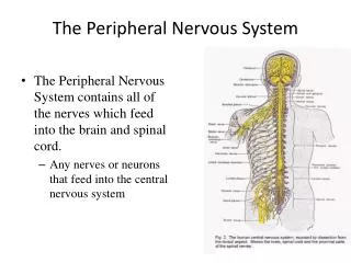
The Peripheral Nervous System. Chapter 14. Introduction to the PNS. Nervous structures outside the brain and spinal cord - sensory and motor connections to the outside world - nerves thread throughout the body to allow the CNS to receive information and take action

The Peripheral Nervous System
E N D
Presentation Transcript
The Peripheral Nervous System Chapter 14
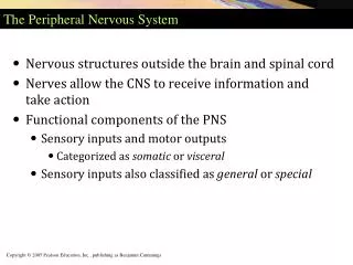
Introduction to the PNS • Nervous structures outside the brain and spinal cord - sensory and motor connections to the outside world - nerves thread throughout the body to allow the CNS to receive information and take action • Functional components of the PNS - sensory inputs and motor outputs - categorized as somatic or visceral - also classified as general or special
Functional Components of the PNS • Basic structural components: 1. Sensory receptors – pick up stimuli from inside and outside the body, then initiate impulses in sensory axons 2. Motor endings – the axon terminals of motor neurons that innervate the effectors 3. Nerves – bundles of peripheral axons and Ganglia - clusters of peripheral neuronal cell bodies - most are mixed nerves, contain both sensory and motor axons - some cranial nerves are purely sensory or purely motor in function
Autonomic Nervous System • General visceral motor part of the PNS • Has 2 divisions (with opposite effects): - Parasympathetic: ‘housekeeping’ activities (rest and digest) - Sympathetic: extreme situations (fight or flight)
Functional Organization of the PNS Figure 14.1
Peripheral Sensory Receptors • Most fit into 2 main categories: 1. free nerve endings of sensory neurons - monitor general sensory information such as touch, pain, pressure, temperature, and proprioception 2. complete receptor cells – specialized epithelial cells or small neurons that transfer sensory information to sensory neurons - monitor most special sensory information such as taste, vision, hearing, and equilibrium
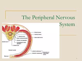
Basic Anatomical Scheme of the PNS in the Region of a Spinal Nerve A nerve is composed of numerous nerve fibers Figure 14.2
Sensory Receptors of the PNS • Also classified according to: a) Location – based on body location or location of stimuli to which they respond b) Type of stimulus detected – kinds of stimuli that most readily activate them c) Structure – divided into 2 broad categories free or encapsulated nerve endings
Classification by Location • Exteroceptors – sensitive to stimuli arising from outside the body - located at or near body surfaces - include receptors for touch, pressure, pain, temperature, and most receptors of the special sense organs • Interoceptors (visceroceptors) – receive stimuli from internal viscera (digestive tube, bladder, lungs) - monitor a variety of stimuli such as changes in chemical concentration, taste stimuli, stretching of tissues, and temperature - activation causes visceral pain, nausea, hunger, or satiety
Classification by Location • Proprioceptors – monitors degree of stretch and sends input on body movements to the CNS - located in musculoskeletal organs such as skeletal muscles, tendons, joints, and ligaments
Classification by Stimulus Detected • Mechanoreceptors – respond to mechanical forces - such as touch, pressure, stretch, vibrations, and itch • Thermoreceptors – respond to temperature changes • Chemoreceptors – respond to chemicals in solution (molecules tasted or smelled) and to change in blood chemistry • Photoreceptors in the eye – respond to light • Nociceptors – respond to harmful stimuli that result in pain (noci = harm)
Classification by Structure • General sensory receptors – widely distributed • Nerve endings of sensory neurons moniter: - Touch - Pressure - Vibration - Stretch - Pain - Temperture - Proprioception
Classification by Structure • General sensory receptors are divided into 2 groups - Free nerve endings - Encapsulated nerve endings Note: there is no perfect ‘one receptor – one function’ - one receptor can respond to several kinds of stimuli and different receptor types can respond to similar stimuli
Free Nerve Endings • Abundant in epithelia and underlying CT • Respond to pain and temperature • Monitor affective senses – those to which people have an emotional response (pain) • 2 specialized types of free nerve endings: - Merkel discs: lie in the epidermis - Hair follicle receptors: wrap around hair follicles
Free Nerve Endings • Merkel discs – a disc-shaped epithelial cell innervated by a sensory nerve ending - slowly adapting receptors for light touch (respond and send out action potentials even after continual stimulation) • Hair follicle receptors – receptors for light touch - monitor the bending of hairs - rapidly adapting (sensation disappears quickly even if the stimulus is maintained) • Itch receptor – in the dermis (newly discovered)
Encapsulated Nerve Endings • Consist of one or more end fibers of sensory neurons enclosed in connective tissue • All seem to be mechanoreceptors – capsules either amplify the stimulus or filter out the wrong types of stimuli • 4 main types - Meissner’s corpuscles - Pacinian corpuscles - Ruffini endings - Proprioceptors
Meissner’s Corpuscles • Spiraling nerve ending surrounded by Schwann cells - occur in the dermal papillae - rapidly adapting receptors for discriminative touch - occur in sensitive, hairless areas of the skin
Meissner’s Corpuscles Table 14.1 (2 of 4)
Pacinian Corpuscles and Ruffini Endings • Pacinian corpuscle - single nerve ending - surrounded by layers of flattened Schwann cells - occur in the hypodermis - sensitive to deep pressure - rapidly adapting receptors • Ruffini endings – located in the dermis - monitor continuous pressure on the skin (adapt slowly)
Proprioceptors • 3 types - monitor stretch in locomotory organs: • Muscle spindles - measure the changing length of a muscle - imbedded in the perimysium between muscle fascicles • Golgi tendon organs – located near the muscle-tendon junction - monitor tension within tendons • Joint kinesthetic receptors – sensory nerve endings within the joint capsules
Structure of Proprioceptors Figure 14.4
Cranial Nerves • Attach to the brain and pass through foramina of the skull • Numbered from I – XII • Cranial nerves I and II attach to the forebrain - all others attach to the brain stem • Primarily serve head and neck structures - except the vagus nerve (X) that extends into the abdomen
The 12 pairs of cranial nerves Figure 14.5
Olfactory Nerves • Sensory nerves of smell Table 14.3 (1 of 12)
Optic Nerve • Sensory nerve of vision Table 14.3 (2 of 12)
Oculomotor Nerve • Innervates four of the extrinsic eye muscles Table 14.3 (3 of 12)
Trochlear Nerve • Innervates the superior oblique muscle (extrinsic eye muscle) Table 14.3 (4 of 12)
Trigeminal Nerve Table 14.3 (5 of 12) • Provides sensory innervation to the face and motor innervation to chewing muscles
Abducens Nerve • Abducts the eyeball – innervates lateral rectus muscle Table 14.3 (6 of 12)
Facial Nerve • Innervates muscles of facial expression Table 14.3 (7 of 12)
Vestibulocochlear Nerve • Sensory nerve of hearing and balance Table 14.3 (8 of 12)
Glossopharyngeal Nerve • Innervates structures of the tongue and pharynx Table 14.3 (9 of 12)
Vagus Nerve • A mixed sensory and motor nerve • “Wanders” into thorax and abdomen • Parasympathetic innervation of organs Table 14.3 (10 of 12)
Accessory Nerve • An accessory part of the vagus nerve • Innervates trapezius muscle Table 14.3 (11 of 12)
Hypoglossal Nerve • Runs inferior to the tongue - innervates the tongue muscles Table 14.3 (12 of 12)
Spinal Nerves • 31 pairs – contain thousands of nerve fibers • Connect to the spinal cord • Named for point of issue from the spinal cord - 8 pairs of cervical nerves (C1 – C8) - 12 pairs of thoracic nerves (T1 – T12) - 5 pairs of lumbar nerves (L1 – L5) - 1 pair of coccygeal nerves (Co1)
Spinal Nerves Posterior View Figure 14.6
Spinal Nerves • Connect to the spinal cord by the dorsal root and ventral root • Dorsal root – contains sensory fibers - cell bodies located in the dorsal root ganglion • Ventral root– contains motor fibers arising from anterior gray column
Spinal Nerves • Branch into dorsal ramus and ventral ramus - dorsal and ventral rami contain sensory and motor fibers • Rami communicantes – connect to the base of the ventral ramus - lead to the sympathetic chain ganglia
Spinal Nerves Figure 14.7a
Innervation of the Back • Dorsal rami – innervate back muscles - follow a neat, segmented pattern - innervate a horizontal strip of muscle and skin (in line with emergence point from the vertebral column)
Innervation of the Back Figure 14.7b
Innervation of the Anterior Thoracic and Abdominal Wall • Thoracic region – ventral rami arranged in simple, segmented pattern • Intercostal nerves supply intercostal muscles, skin, and abdominal wall - each gives off lateral and anterior cutaneous branches
Introduction to Nerve Plexuses • A network of nerves • Ventral rami (except T2 – T12) - branch and join with one another - form nerve plexuses in the cervical, brachial, lumbar, and sacral regions - primarily serve the limbs - fibers from ventral rami crisscross
The Cervical Plexus • Buried deep in the neck under the sternocleidomastoid muscle • Formed by ventral rami of first 4 cervical nerves (C1 – C4) • Most are cutaneous nerves • Some innervate muscles of the anterior neck • Phrenic nerve – major nerve
The Brachial Plexus and Innervation of the Upper Limb • Brachial plexus lies in the neck and axilla • Formed by ventral rami of C5 – C8 • Cords give rise to main nerves of the upper limb Fig 14.9d
Nerves from Lateral and Medial Cords • Musculocutaneous – main branch of the lateral cord - innervates the biceps brachii and brachialis • Median – originates from both lateral and medial cords - innervates anterior forearm muscles and lateral palm • Ulnar – branches from the medial cord - innervates intrinsic hand muscles and skin of the medial hand • Radial – continuation of the posterior cord - largest branch innervates muscles of posterior upper limb • Axillary – innervates the deltoid and teres minor