THE BLOOD
THE BLOOD. The 3 Main Functions of Blood :. Transportation Protection Regulation Blood is a connective tissue in liquid form Greatest benefit from homeostasis Continuous flow of blood thru 60,000 miles of blood vessels . TRANSPORTATION :. Blood moves thru body where cells receive:
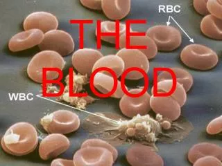

THE BLOOD
E N D
Presentation Transcript
The 3 Main Functions of Blood: • Transportation • Protection • Regulation • Blood is a connective tissue in liquid form • Greatest benefit from homeostasis • Continuous flow of blood thru 60,000 miles of blood vessels
TRANSPORTATION: • Blood moves thru body where cells receive: • Nutrients from digestive organs • Oxygen from lungs • Hormones secreted from endocrine gland • Cells give blood waste • (CO2, urea & uric acid) & their secretions
Protection: • From harmful microorganism & their toxins • Through Phagocytic white blood cells • Specialized proteins called Antibodies • Against fluid loss after an injury by clotting
Regulation: • Regulates acid-base balance of the body fluids • By way of buffers • Neutralize potential harmful effects of: • too much CO2 • lactic acid • other compounds • Body temp. by cooling or heating parts of body • Controlled by Hypothalamus • Controls volume of blood flow to diff. areas of body Figure This figure highlights some of the major acute (short-term) effects on the body during exercise.
Properties of Blood: • Color • Volume • pH
COLOR • RED COLOR = • HEMOGLOBIN (PIGMENT PROTEIN) • Arterial blood the O2 molecules are chemically bound to hemoglobin • Crimson-red color • Venous blood O2molecules are not as prevalent & blood= • Dark red color w/a slightly bluish tint • SEEN THROUGH SKIN VEINS LOOK GREENISH- BLUE but it is NOT GREEN OR BLUE
VOLUME • 8% OF BODY WEIGHT • Most in vessels--rest in heart • Does not vary much from day to day or year to year • Avg. Male = • 5-6 liters of blood • Avg. Female = • 4-5 liters of blood • Difference due to avg body weight not sex Apx. 8 pints
Blood is thicker, denser, & more adhesive than H2O • Due to formed elements (red blood cells) • Causes blood to flow 5x slower than H2O • Resistance to flow = viscosity • Therefore blood is a viscous substance b/c it resists flow more than water Figure The shear rate dependence of normal human blood viscoelasticity at 2 Hz and 22 °C.
pH • Slightly alkaline (aka: basic) • pH = 7.35-7.45 • Range stays small despite change in: • Diet • Cell secretions • Metabolic rateby buffering systems that remove H+ ions • If buffers fail: • BLOOD TOO ACIDIC (pH below 6.0) • Body cells stop functioning • No homeostasis = Acidosis • Too little acid in blood = Alkalosis (a lot less common) • Possible complications = Arrhythmias, Coma, Electrolyte imbalance (such as hypokalemia)
Leukocytes • Less than 1% of total blood volume • 5000 TO 10,000 in cubic mm • Any change in number… • High or low indicates a disease
Types: • All contain a nucleus • (unlike the RBC’s) • Can wander outside the Circ. System- Diapedesis • WBC cells differ in: • Nature of cytoplasm • Size • Shape of nucleus • Response to different staining techniques • Divided into 2 groups by cytoplasm differences: • Granulocytes • Agranulocytes STOP HERE
Granulocytes • Cytoplasm contains highly visible pebble-like objects, known as granules • Twice the size of RBC’s • They contain a nucleus that is split into sections called lobes • Produced in red marrow
Three types: • Eosinophils • Neutrophils • Basophils • Names come from the type of stain that brings out their distinguishing features→ • Neutral • Eosin • Basic
Neutrophil: • Most abundant = granulocyte • Stain pink in a neutral stain • Nucleus contains: 2 to 5 lobes→ • Interconnected by thin bridges • Make up about 60% of all WBC’s in a normal blood sample
Eosinophils: • 1 to 4% of WBC’s in a normal blood sample • Granules stain red in an acid stain that contains a dye known as eosin • Nucleus = 2 lobes
Eosinophils are: • Not very mobile • Not very active • Can phagocytize certain foreign particles produced by allergic reactions → • Invading parasites • Pollen grains • Mold spores
Basophils: • Most rare→0.5% or less of WBC’s in blood • Large granules that stain blue in basic stain • Nucleus is often bent into an s-shape with 2 lobes
Basophils & Mast cells produce a substance called = histamine • causes swelling or inflammation • Swelling tells other WBC’s where to find the site of infection ***Mast cells reside in tissues in the body, and basophils are in the blood stream. http://link.brightcove.com/services/link/bcpid236059233/bctid347806799
Agranulocytes: • Contain very small amount of cytoplasmic granules • 2 types of cells → • Monocytes • Lymphocytes • Both produced in red bone marrow • Also produced by organs of lymphatic system → • Lymph nodes • Spleen • Thalamus
Monocyte: • Largest cells in blood • 3x larger than RBC’s • 2x larger than granulocytes • Nucleus can be round, oval, or lobed • Often occupies most of the cell volume • 3 to 8% of WBC’s in a blood sample
Lymphocyte: • Same size as the RBC = the smallest WBC • Nucleus is round and large → • Takes up almost all of cell volume • 25-33% of WBC’s in a blood sample
Function: • Protection from disease • Move out of vessels = diapedesis • Once in the intestinal fluid they act like amoeba, extending streams of cytoplasmic arms called = pseudopodia • To find infection they sense chemicals released by invading microorganisms & damages cells
Once found the WBC traps the microorganism and engulfs it = phagocytosis • The primary cells used for phagocytosis ar the neutrophils & monocytes • Neutrophils are mobile & usually arrive 1st at site of infection • Monocytes are very active too, large size allows for phagocytizing whole cells & large # of bacteria
When more WBC’s arrive at the site of infection they form a collection of living—dead—broken cells and plasma = pus • Not only phagocytosis to combat disease: • Highly specific proteinsproduced by the lymphocytes = • Antibodies → • These act against foreign particles and toxins that enter body • Production of antibodies = immunity http://gu.com/p/2kntv
Platelets: • Aka → Thrombocytes • Formed elements that are fragments of complex cells • During development in red bone marrow, they are formed when a large precursor cell breaks apart • In small fragments platelets are released into blood stream for circulation
Larger fragments are broken down further to form more platelets • Each platelet contains: • Cytoplasm surrounded by a plasma membrane • No nucleus but most organelles found in cytoplasm • 1/10 the size of a RBC • Shape = round or oval disk
150,000 to 360,000 platelets per cubic mm in normal blood sample = less numerous than RBC • Prevention of fluid loss • Initiate the formation of blood clots • This plugs up the breaks in the blood vessel wall after an injury