Download
1 / 1
20 likes | 246 Views
Motor areas. Tumor. Tumor. Visual cortex. Tumor. Motor Activity. Teaching points. The cause of a pause is not always cardiac. References.

E N D
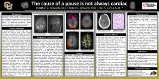
Motor areas Tumor Tumor Visual cortex Tumor Motor Activity Teaching points The cause of a pause is not always cardiac References While state-of-the art technology and cutting-edge diagnostic tools aid in diagnosis, thorough history (persistent “dizzy spells” in this case) and physical exam still reign supreme in patient care The search for secondary – or even tertiary – causes must be considered in diagnosis; in this case the secondary cause was seizures leading to asystole, and the tertiary cause being tumor MRI may be performed with traditional pacemakers, utilizing low-energy protocol sequences and close cardiac monitoring fMRI has markedly changed and improved neurosurgical resection of malignancy, specifically improving outcomes by avoiding critical speech, motor, and vision pathways DTI can identify specific neuronal tracts by imaging water molecule diffusion The general public should be trained in BLS/CPR, as immediate and effective resuscitation likely saved this patient’s life Contact information Jonathan Schwartz, M.D. University of Colorado Denver Internal Medicine Residency Program jgschwartz@yahoo.com Literature Review GBM is the most common and most malignant glial tumor, 60% of all primary brain cancers Median survival is 14 months after diagnosis One study reported 5/1244 (0.4%) of patients with epilepsy suffered ictal asystole While seizures are common in patients with brain tumors, they are less common with high-grade compared to low-grade gliomas Patients with brain tumors in the motor cortex are at higher risk of having seizures GBM is a relatively rare cause of temporal lobe seizures; 12% of cases in one series Bradycardia, SA node dysfunction, and asystoleonly rarely present as the first sign of cerebral malignancy Ictal asystole is a rare but often fatal seizure complication, and is an important cause of sudden unexplained death in epilepsy (SUDEP) Ictal asystole is most commonly associated with temporal lobe seizures but has also been documented with frontal lobe seizures New data suggest treating the underlying cause of epilepsy may eliminate the need for pacemaker implantation Imaging Jonathan G. Schwartz, M.D.1, Robert S. Schwartz, M.D.2, Joel A. Garcia, M.D.1,3 1. Seeck, M., et al. Symptomatic postictal cardiac asystole in a young patient with partial seizures. Europace 2001 Jul;3(3):247. 2. Devinsky, O. Bradycardia/asystoleinduced by partial seizures: a case report and review. Neurology 1997 Jun;48(6):1712-4. 3. Van der Sluijs B.M., et al. Brain tumor as a rare cause of cardiac syncope. J of Neurooncology 2004 Mar-Apr;67(1-2):241-4. Background Although brain MRI was indicated, it could not be performed because of the recent pacemaker implant. Contrast head CT was substituted, and suggested a vague temporoparietal lesion. MRI was now mandated, and a low-energy MRI protocol was used with temporary pacemaker reprogramming. A cardiologist was present throughout the scan. MRI showed a high-grade glioblastoma multiforme (GBM, Images 1 and 2), which raised concern for temporal lobe seizures as a cause of the lightheaded amnestic episodes. Furthermore, temporal lobe seizures became a possible explanation of his asystole. Consultation with an epileptologist and subsequent EEG confirmed temporal lobe seizures. The patient was initiated on antiepileptic drug therapy. The implanted pacemaker was removed and functional MRI (fMRI) was performed to determine if the tumor could be removed without impacting key vision, speech, and motor control centers. The tumor was successfully removed and GBM pathologically confirmed. An investigational, MRI-compatible pacemaker was implanted since the patient would need multiple follow-up MRI scans. MRI showed a high-grade temporoparietalGBM (Images 1 and 2). fMRI imaging was performed to plan safe tumor removal. fMRI during reading aloud (Image 3) shows close tumor proximity to the eloquent cortex. Tongue tapping (Image 4) and left finger tapping (Image 5) images indicate surgical resection was possible without significant postoperative deficit. Diffusion tensor imaging (Image 6) shows a displaced right optic radiation without tumor interruption. Contrast head CT (Image 7) shows poor tumor visualization compared to MRI. Tumor histopathology (Image 8) showed typical GBM with heterogeneous mixtures of poorly-differentiated neoplastic astrocytes. 1Department of Medicine, University of Colorado Denver, Aurora, CO; 2Minneapolis Heart Institute, Minneapolis, MN; 3Division of Cardiology, University of Colorado Denver, Aurora, CO Workup and clinical outcome Syncope is a frequent cause of hospital admission, and cardiac causes must be considered and ruled out. Cardiogenic syncope results from sinoatrial or atrioventricular node disease, conduction system disease, or ventricular arrhythmias. Rarely, however, cardiogenic syncope has secondary or even tertiary causes, as this case illustrates. Image 8: Histopathology of glioblastomamultiforme A previously-healthy 49 year old male was referred for follow-up after a dual chamber pacemaker was implanted one week prior. He was on vacation and was crossing the street when he suddenly lost consciousness. His wife, a critical care nurse, could find no pulse and immediately began CPR. The patient developed a pulse and regained consciousness. EMS arrived, placed him on continuous monitoring, and he again lost pulses and suffered a generalized seizure. He again regained pulses and was taken to a nearby ED for evaluation. Initial ECG revealed extreme bradycardia (see rhythm strip above) followed by sinus arrest, and he again lost consciousness. He was revived and again regained consciousness. Head CT, echocardiography, and metabolic panel were unremarkable. A temporary pacemaker was inserted, followed by permanent pacemaker implant. He was discharged home, but had persistent “lightheaded” and “dizzy” episodes for which he presented to our emergency department. He was referred to our clinic for pacemaker interrogation, which showed normal function. A careful history revealed these lightheadedness episodes were frequent, and associated with amnesia. This was concerning, especially since they persisted despite normal cardiac and pacemaker function. This warranted further workup, with particular focus on neurologic evaluation. ECG rhythm strip obtained upon EMS arrival Image 1: Transverse MRI with temporoparietal lesion Image 2: Sagittal MRI with temporoparietal lesion Image 3: Transverse fMRI, story reading Image 4: Transverse fMRI, tongue tapping Image 5: Transverse fMRI, left finger tapping Image 6: Diffusion Tensor Imaging with optic radiations Image 7: Contrast CT showing poor discrimination of tumor Initial presentation Tumor Visual tracts


