Download
1 / 1
10 likes | 96 Views
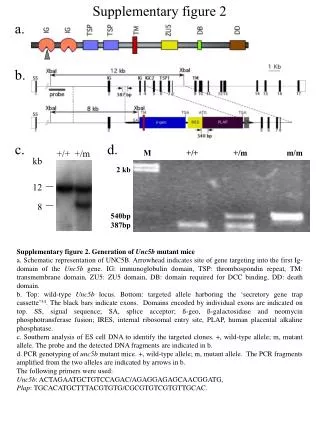
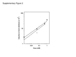
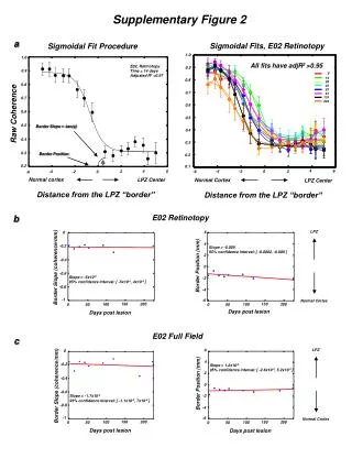
Explore retinotopy in normal cortex post-lesion, days 100-200, using sigmoidal fits with adjusted R2>0.95. Assess LPZ center- border coherence, position, and slope for detailed mapping. Investigate LPZ distance and effects of post-lesion time on retinotopy adjustments with 95% confidence intervals. Analysis performed for Supplementary Figure 2a.

E N D
Sigmoidal Fits, E02 Retinotopy All fits have adjR2 >0.95 1.0 1.0 0.9 0.9 0.8 0.8 0.7 0.7 0.6 0.6 0.5 0.5 0.4 0.4 0.3 Normal Cortex LPZ Center 0.3 0.2 0.2 0.1 4 4 6 6 2 2 -4 -4 -2 -2 0 0 -6 -6 0 14 28 42 53 91 119 224 Distance from the LPZ “border” Normal cortex LPZ Center E02 Retinotopy LPZ 6 6 0 0 4 4 -0.2 -0.2 Slope = -0.005 95% confidence interval: [ -0.0002, -0.009 ] 2 2 -0.4 -0.4 Border Position (mm) Border Position (mm) Border Slope (coherence/mm) Border Slope (coherence/mm) 0 0 -0.6 -0.6 Slope = -5x10⁻⁵ 95% confidence interval: [ -5x10⁻⁴, 4x10⁻⁴ ] -2 -2 -0.8 -0.8 -4 -4 -1 -1 -6 -6 Normal Cortex Normal Cortex Days post lesion Days post lesion 150 150 150 150 200 200 200 200 100 100 100 100 0 0 0 0 50 50 50 50 E02 Full Field c LPZ Slope = 1.2x10⁻³ 95% confidence interval: [ -2.6x10⁻³, 5.2x10⁻³ ] Slope = -1.7x10⁻⁴ 95% confidence interval: [ -1.1x10⁻³, 7x10⁻⁴ ] Days post lesion Days post lesion Supplementary Figure 2 a Sigmoidal Fit Procedure E02, Retinotopy Time = 14 days Adjusted R2 =0.97 Raw Coherence Border Slope = -tan(f) Border Position f Distance from the LPZ “border” b