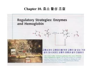
Chapter 10. 효소 활성 조절
Chapter 10. 효소 활성 조절. 교통순경이 교통정리를 하면 교통이 잘 되는 거와 같이 대사과정도 교통의 흐름과 같이 조절된다. 요 점. ⎕ Principal four regulatory ways of enzymatic activities. 1. Allosteric control. 1) Aspartate transcarbamoylase (ATCase): feed-back inhibition. 2) Hemoglobin (Hb): 산소결합은 협동적 , H + , CO 2 , 2,3-BPG 에 의해 조절.

Chapter 10. 효소 활성 조절
E N D
Presentation Transcript
Chapter 10. 효소 활성 조절 교통순경이 교통정리를 하면 교통이 잘 되는 거와 같이 대사과정도 교통의 흐름과 같이 조절된다
요 점 ⎕ Principal four regulatory ways of enzymatic activities 1. Allosteric control 1) Aspartate transcarbamoylase (ATCase): feed-back inhibition 2) Hemoglobin (Hb): 산소결합은 협동적, H+, CO2, 2,3-BPG에 의해 조절 2. Multiple forms of enzyme (isozyme) - 조직, 장기 등에 분포; 동일 반응 촉매; - 구조, Km, Vmax 등에서 차이 3. Reversible covalent modification - 인산화와 탈인산화에 의한 조절 4. Proteolytic activation -Zymogens or proenzymes: 활성화 불활성화 -Chymotrypsinogen, proelastase, procaspase, blot clotting
I. Aspartate transcarbamoylase (ATCase) : Allosteric control 1) Feed-back inhibited by the final product (CTP) of the pyrimidine pathway. - CTP bind to regulatory site (not active site) or allosteric site
2) Consists of separable catalytic and regulatory subunits -PHB 처리로 분리됨 (C6 + R6) (A) 자연상태의 ATCase (B) PHB으로 효소를 처리한 후
3) Allosteric interactions are mediated by large changes in quaternary structure
4) Allosterically regulated enzymes do not follow Michalis-Menten kinteics
II. LDH(Isozymes) H isozyme (heart에 발현: 호기성); M isozyme (골격 근육에서 발현: 혐기성) -aa 서열이 75% 동일 -H4: heart에서 발견, 기질과 친화력이 강 (M4보다) -H3M: 두 가지 사슬들의 비에 따라 중간의 성질들을 가진다. A : 쥐 심장의 LDH의 동질효소들의 조성 은 발생과정 동안 변한다. H동질효소를 네모로 그리고 M동질효소를 원으로 나타내었다.부의 수와 정의 수는 각각 태어나기 전후의 날수를 가리킨다. B : LDH의 동질효소들의 함량은 조직이 다르다.
1) Phosphorylation is a highly effective mean of switching the activity of target proteins (1) Tyr or ser 잔기 인산화 (inside cells) (인산화가 일어나는 6가지 이유) ①인산기는 변형된 단백질에 두 개의 음전하를 더한다. ②인산기는 세 개 또는 세 개 이상의 수소결합을 형성할 수 있다. ③인산화 반응의 자유에너지 변화는 크다. ④인산화와 탈인산화는 1초보다 짧은 시간에 일어날 수도 있고, 수시간 동안 일어날 수도 있다. ⑤인산화는 크게 증폭된 효과들을 이끌어 내는 일이 흔히 있다. ⑥ATP는 세포에서의 에너지 유통 매체이다. (2) 주요 protein kinases (Table 2)
(3) cAMP activates protein kinase A (PKA) by unleashing its catalytic subunits 네 분자의 CAMP의 결합은 억제된 완전효소(R2C2)를 조절 소단위체(R2)와 두 개의 촉매 활성을 가진 소단위체들(C)로 해리시킴으로써 단백질 키나아제 A를 활성화 한다.
IV. Many enzymes are activated by specific proteolytic cleavage
1) Chymotrypsin (1) Activated by specific cleavage of a single peptide bond -키모트립신의 세 사슬들은 두개의 사슬간 이황화결합으로 연결된다. α (2) Activation leads to the formation of a substrate binding site 활성 미모트립신의 구조에 필수적인 아스파르트산 194의 카르복실기와 이소루신 16의 아미노기 사이의 정전기적 상호작용은 키모트립신에서만 가능하다.
(3) Trypsin은 다른 zymogen을 활성화 시킴 - 엔테로펩티드 가수분해효소는 트립신을 활성화함으로써 췌장에서 분비되는 지모겐들의 활성화를 개시한다. - 트립신은 다른 지모겐들을 활성화한다. - 활성 효소들은 노란색으로 나타내었고, 지모겐들을 주황색으로 나타내었다.
(4) Pancreatic trypsin inhibitor binds very tightly to the active site of trypsin - 트립신(노란색)과 췌장의 트립신 억제물(빨간색)의 복합체의 구조. - 억제물의 리신15는 효소의 활성자리 안으로 들어가서 활성자리에 있는 아스파르트산 189와 염다리결합을 형성한다. - 결합된 억제물과 유리된 억제물 들은 구조가 거의 동일하다.
2) Clotting occurs by a cascade of zymogen activations (1) Cascade 활성화
(2) Prothrombin은 vitamin k 에 의해 thrombin으로 전환 첫번째 영역을 글라 영역(gla domain)이라 부르고 영역 2와 3을 크링글 영역들(kringle domain)이라고 부른다. - Vitamin K is required for the synthesis of prothrombin and other calcium-binding proteins 비타민 K-의존성 카르복실화반응은, Ca2+의 약한 킬레이트제인 글루탐산을 훨씬 더 강한 킬레이트제인 γ –카르복시글구탐산으로 변환 시킨다.
(3) Fibrinogen is converted by thrombin into a fibrin clot (A) 리본그림이다. 두 막대 구역들은 α–나선형 감긴코일이며 각각의 말단에서 구상 구역에 연결되어 있다. (B) 피브리노펩티트 A와 B의 위치들을 나타내는 모식도이다.
(4) Fibrin clots are lysed by plasmin - 트롬빈이 피브리노겐의 중앙의 구로부터 피브리노펩티드 A외 B를 쪼갠다. - β 와 γ사슬들의 카르복실-말단에 있는 구형 영역들이 β 와 α사슬들의 아미노- 말단에서 노출된 “마디들”과 상호작용하여 덩어리들을 형성한다.