Download
1 / 46
460 likes | 488 Views
This presentation covers the basics of renal system imaging modalities, anatomy, and illustrative cases, aided by Dr. Dima's notes in dark red. From conventional radiography to nuclear medicine, explore the world of kidney imaging. Master the terminology and understand key features of each imaging modality. Excel in diagnosing renal pathologies and enhancing patient care with this informative guide.

E N D
Imaging of the Renal System Dr. DimaJamjoom Department of Radiology Same slides but with Dr.Dima’s notes. (Dark Red) Good luck اجتهاد شخصي إن أصبت فمن الله وان أخطأت فمن نفسي
OUTLINE • Introduction • Imaging modalities • Anatomy • Cases
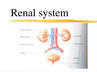
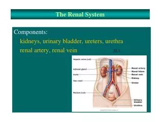
INTRODUCTION • What is radiology? It is a medical specialty that employs the use of imaging to both diagnose and treat disease within the human body. • What is the renal system? Kidneys, ureters, urinary bladder and urethra.
IMAGING MODALITIES • Conventional radiography • Intravenous urogram (IVU) • US • CT • MRI • Nuclear medicine
Terminology This slide is *taken from the boys slide*, I think it’s the only difference between the Female/Male slides. • X ray : Radio-opaque (white) vsradio-lucent (black) • US: Hyper-echoic (white) / hypo-echoic(black) • CT: Hyper-dense (white) / hypo-dense(black) • MRI: Hyper-signal (white) / hypo-signal • Nuclear med.: Highuptake(black)/ lowuptake(white)
Conventional radiography= Plain X-Ray • First imaging modality.meaning that a patient complains of symptoms renal disease, we start by X-ray • Cheap. • Useful for radio-opaque stones. But it’s limited, usually used only for this purpose. *radio-opaque stones will appear white.* Note that there are stones which are not radio-opaque and are diagnosed by US and CT.
Conventional radiography Image features: • Projectional image. • Image contrast determined by tissue density. • Good evaluation radio-opaque stones.
IVU= same as plain x-ray but we give the patient contrast • Conventional x-ray plus intravenous contrast. • Cheap. • Recently replaced by CT and MRI. • Useful for radio-opaque stones. Contrast : مادة نحقنها عبر الوريد + it will appear white in the x-Ray.
IVU Image features: • Projectional image. • Image contrast determined by tissue density and IV contrast. • Good evaluation of collecting system and radio-opaque stones. + the doctor said it is used also for hydronephrosis which is dilatation of the collecting system.
US • Use high frequency sound wave. • Contrast between tissue is determined by sound reflection.
US Image features: • Operator dependant. Meaning that if the practitioner is good, then images will be good if not then the images will be bad. • Projectional image. • Good resolution. • Used for stone, hydronephrosis, focal lesion. Fluid = black. Fat = White.
CT • Same basic principle of radiography. • More precise. • Costly. • +/- contrast.( we can do it with or without contrast) • Useful for trauma, stone, tumor, infection. If patient suspected with stones, then the gold standard is CT without contrast.
CT Non-contrast CT Image features: • Cross sectional images. • Image contrast determined by tissue density +/- contrast. • Better evaluation of soft tissue.
MRI • Better evaluation of soft tissue. • Expensive. + time consumer, takes 30-45 mins • Useful for soft tissue pathology: tumor, infection. If we are suspecting renal tumors, then MRI is the best.
MRI Image features: • Cross sectional images. • Image contrast determine by tissue properties. • Excellent for soft tissue evaluation.
Nuclear medicine • Utilizes a gamma camera and radioactive isotopes*. • Functional test. Meaning that the Anatomy won’t appear clear as the CT and MRI but gives us an idea about the function of the kidneys. • Less expensive. • Useful for: obstruction and split function. * مادة مشعة نحقنها عبر الوريد
Nuclear medicine Image features: • Projectional image. • Image contrast by tissue uptake and metabolism.
Plain x-ray Kidneys size is about 3 and half vertebral bodies including the vertebral discs.
CT The doctor talked in details about the fasciae and everything in this picture.
CT with contrast , • *Aorta* : we can see it in the right because we gave contrast.
What are the imaging modalities? • What are the findings? • Diagnosis?
Case (1) • Young male patient presenting with left flank pain and hematuria, no fever and normal WBC count.
This is IVU we can see dilatation of the collecting system ( hydronephrosis).. Stones causing hydronephrosis. *Renal pelvis is a bet dilated* This is Plain X-ray without contrast, we can see radio-opaque stones in the left kidney.
Note the dilatation on this side ( Hydronephrosis ) Same patient but with CT and US, stones will be white by ultrasound.
Case (2) • Middle aged woman presenting with flank pain, fever and high WBC. We expect infection.
CT Kidneys are white except some areas which is darker, this is the focal infection ( pyleonephritis )
Case (3) • Elderly male patient with recurrent urinary tract infections.
Renal pelvis is dilated. * it’s dark because it contains fluid* us
Nuclear, collecting system is dilated. (normally the isotope will be less in the kidneys with time) but in this case still there is isotope in the kidneys.
Case (4) • Young female presenting with decreased renal function (high urea and creatinine level). Renal failure.
US • Kidneys are not seen, it’s replaced by cysts which are dark. This is congenital ( autosomal dominant polycystic kidney disease.) MRI of same patient. No kidneys, replaced by cysts.
Case (5) • Elderly male patient with painless hematuria and weight loss. tumor
Case (6) • Young male patient involved in a motor vehicle accident with blunt trauma to the abdomen. Trauma, cases of trauma = CT
Renal trauma grading bleeding beneath the renal capsule, contusion. (دائرة) Ulceration less than 2 cm + not involving the collecting system ( ألسر = طويل شوي مو دائرة ) more than 2 cm but also not involving the collecting system Extending to collecting system or if there is( thrombus ) , injury to blood supply. Shattered kidney, multiple injuries , or اذا انقطع البلود سبلاي تماما
References • Stephanie Ryan, “Anatomy for Diagnostic imaging”, 2nd Edition. • Jamie Weir, Peter Abraham, “Imaging Atlas of Human Anatomy”3rd Edition. • Peter Armstrong, “diagnostic imaging”, 5th Edition.