Download
1 / 46
520 likes | 978 Views
RADIOLOGY OF THE RENAL SYSTEM. DR. Reshaid Aljurayyan DEPARTMENT OF RADIOLOGY. Outline:. Introduction Imaging modalities used to study the renal system Anatomy and normal appearance of the renal system Common pathological cases. Introduction. What is the radiology?

E N D
RADIOLOGY OF THE RENAL SYSTEM DR. Reshaid Aljurayyan DEPARTMENT OF RADIOLOGY
Outline: • Introduction • Imaging modalities used to study the renal system • Anatomy and normal appearance of the renal system • Common pathological cases
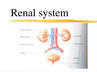
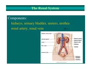
Introduction • What is the radiology? Radiology is a medical specialty that employs the use of imaging to both diagnose and treat disease visualized within the human body. • What is the renal system?
Outline: • Introduction. • Imaging modalities used to study the renal system. • Anatomy and normal appearance of the renal system. • Common pathological cases.
What are the radiological modalities that can be used to image the renal system ?
Imaging modalities: • Conventional X-Ray • IVU (intra-venous urogram) / X-Ray + Contrast • US • CT • MRI • Nuclear medicine
Imaging modalities: • Conventional X-Ray • IVU (intra-venous urogram) / X-Ray + Contrast • US • CT • MRI • Nuclear medicine ALL CAN BE USED
Conventional Radiography (X-ray) • +ve: • Cheap & widely available • Often used as first choice • -ve: • Radiation • Limited anatomy • Used for: • Evaluate abdomen pain • Some time good for diagnosing kidney stones
Radio lucent black (air) • Radio opaque white (bone/stone)
Radiograph (X-ray) Where are the kidneys ??
Radiograph (X-ray) Where are the kidneys ??
IVU • Same as X ray but with IV contrast
IVU • +ve: • Cheap & available • -ve: • Radiation • Needs IV contrast (?reaction) • Old (replaced by CT & MRI) • Used for: • To diagnose kidney stones • To diagnose hydronephrosis
US • Ultrasound. • Use high frequency sound wave. • Contrast between tissue is determined by sound reflection.
US • + ve: • Available • No radiation • Good anatomy • - ve: • Operator dependent • Used for: • Good for kidney stones • Excellent for hydronephrosis • Excellent for focal lesion e.g. cysts, masses
Hyper-echoic white • Hypo-echoic grey • An-echoic black (fluid)
CT: • + ve: • Relatively available (more then MRI) • Very good anatomy • - ve: • Radiation • Some times need IV contrast (? reaction) • Used for: • Excellent for kidney stones (the best) • Excellent for hydronephrosis & masses • Excellent for kidney trauma
Hyper-dense white (stone/bone) • Hypo-dense grey to black (fat/fluid)
MRI • + ve: • Excellent anatomy details • No radiation • - ve: • Expensive • Long scanning time (30 to 60 min) • Not used to diagnosed kidney stone • Used for: • Excellent for masses • Good for hydronephrosis
Hyper-intense (white) • Hypo-intense (grey to black)
Nuclear medicine • + ve: • Excellent to assess function • - ve: • Radiation • Poor anatomy details • Used for: • Evaluated function • Evaluated obstruction
Nuclear medicine • Image features: • Projectional image. • Image contrast by tissue uptake and metabolism.
Objectives: • Introduction. • Imaging modalities used to study the back. • Anatomy and normal appearance of the back. • Common pathological cases.
Case one: • Young male patient presented with left flank pain and hematuria no fever and normal WBC count.
Case two: • Middle age women complaining of flank pain , fever and high WBC.
Case three: • Old male patient complaining of recurrent renal infection.
Case four: • Young female presented with decrease renal function (high urea and creatinine level).
Case six: • old male patient presented with pain less hematura and weight loss.
Case seven: Young male patient involved in road traffic accident with blunt trauma to the abdomen.