Download
1 / 30
600 likes | 1.23k Views
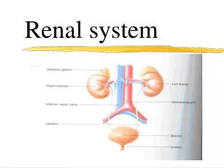
Renal system. By: Ruby zapien, Zulema Vazquez, Sabrina Montano Per. 5. Overview. The urinary system consists of the kidneys, ureters, urinary bladder, and urethra . Aldosterone and antidiuretic hormone (ADH) are important hormones

E N D
Renal system By: Ruby zapien, Zulema Vazquez, Sabrina Montano Per. 5
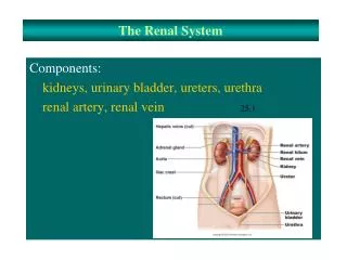
Overview • The urinary system consists of the kidneys, ureters, urinary bladder, and urethra. • Aldosterone and antidiuretic hormone (ADH) are important hormones • Nephrons remove wastes from the blood and regulate water and electrolyte concentrations. • Urine is the product of these functions.
Kidney • Bean-shaped organs • high on the posterior wall • positioned posterior • held in place by adipose tissue and connective tissue • ureter expands into the renal pelvis • renal papillae project into the renal sinus • kidney tissue is divided • Remove metabolic wastes • also help regulate
Kidney Components • Renal Sinus (hilum) • Renal Pelvis • Renal Papillae • Renal Medulla (inner medulla) • Minor Calyces • Renal Cortex (outer cortex) • Renal capsule
Ureters • Narrow • muscular tubes • cross the common iliac arteries • enter the back side of the bladder • transports urine from the kidney to the bladder. • Consists of three layers: -mucous coat -muscular coat -Fibrous coat
Urinary Bladder • Detrusor muscle • Trigone • Ureter • Ductus deferens • Seminical vesicle • Opening of the ureters • Prostate gland • Internal urethra sphincter • Region of external urethral sphincter
Urethra • Passageways that conduct urine from the bladder • Female: short in female • about 1 ½ to 2 inches long • opens into the vestibule of the vagina • Urine only passes though the urethra in females. • Males: long in male • about 6 to 7 inches • From the bladder • Urine and semen passes the urethra
Urethra components • Urethral glands • External Urethral orifice (urinary meatus) • Male (divided into three sections): -Prostatic urethra -membranous urethra -penile urethra
Nephrons • Over one million nephrons in each kidney • A microscopic blood filtering unit • found in the cortex • functional unit of the kidney • Filters blood plasma • absorbs water and salts • secretes unwanted substances
Nephron Components • Renal Corpuscle • Bowman's Capsule (glomercular) • Proximal Convoluted Tubule • Collecting Duct (collecting tubule) • Glomercular Filtrate
Nephron components (cont.) • Juxtaglomercular cells • Juxtaglomecular apparatus • Cortical nephrons • Juxtamedullarynephrons • Podocytes
Nephron Components (cont.) I. Capillaries: • Glomerulus • Afferent arterioles • Peritubular capillary system • Vasa recta • Efferent arterioles
Vasculature: Blood vessels • Renal Arteries • Interlobular arteries • Afferent arterioles • Venous blood • Renal Vein
Vasculature pathway and role • Renal arteryinterlobar artery arcuatearteryinterlobar artery afferent arteriole glomerular capillary efferent arteriole vasa recta and pertiubular capillary interlobular vein arcuate vein arcuate vein interlobular vein renal vein
How urine is created • Created in the nephrons • Filtration, reabsorption, and secretion
filtration • takes place in the glomerular capillaries located in the Bowman’s capsule of the nephrons • This process filters the plasma of the blood, removing wastes and other excess substances and fluids
Filtration rate • directly proportional to the net filtration pressure • If osmotic pressure in glomerulus decreases, the rate drops. If hydrostic pressure in glomerular capsule increases, the rate rises • Auto regulation is a process in which the glomerular filtration rate stays constant
reabsorption • A selective process in which most of the filtered substances gets reabsorbed back into the blood. • Substances pass from the tubules of the nephron into the blood of the peritubular capillaries. • Active transport, endocytosis
Sodium and water reabsorption • Water reabsorption occurs through osmosis and must happen at the proximal convoluted tubule. It can be reabsorbed in the loop of henle. • The reabsorption of water depends on the reabsorption of sodium at the proximal convoluted tubule. • Active transport continues to reabsorb sodium ions through the nephron loop, the distal convoluted tubule, and the collecting duct.
pressure • In filtration, blood pressure forces substances from the capillary into the capsule. • Capsular pressure • colloidal osmotic pressure • Net filtration pressure is the net effect of all the opposing forces of filtration • In reabsorption, adding more fluid to blood vessels increases pressure.
secretion • tubular secretion transports certain substances from the plasma to the tubular fluid. • Various organic compounds and hydrogen ions are actively secreted. • Potassium ions secreted in convoluted tubule and collecting duct.
Regulation • Urine not only eliminates cellular waste products • Our body producing urine • The kidney tubule regulation of the salt and water in our bodies are important factors • Too much or too little water and salt in our bodies can be dangerous. • the urine volume- is adjusted • large amounts of fluid passes into the kidney tubules. • water is being reabsorbed • controlled by a hormone • ADH is secreted • dilute urine is produced • countercurrent mechanism • distal convoluted tubule and collecting duct are impermeable to water • ADH increases the permeability of the distal convoluted tubule and collecting duct
Urine • Urine is the body’s primary waste source. • What is it made out of? • 95% water • Urea • Uric acid • Trace amino acids • Electrolytes
Renal Clearance • The rate at which particular chemical is removed from plasma which indicates kidney efficiency. • 3 renal clearance test to calculate GFR ( Glomerulus filtration rate )
Inulin Clearance Test • Uses a complex polysaccharide. • An amount of insulin is infused into blood at constant rate. • Passes freely through glomerular membranes. • Concentration in the glomerular filtration equals that in the plasma. • Renal tubule not reabsorbed or secreted. • The rate at which inulin appears in urine can be used to calculate the rate of glomerular filtration.
Creatinine Clearance Test • Kidneys remove creatinine from blood. • Creatinine is filtered, but neither reabsorbed nor secreted by kidneys. • Advantage- blood stream normally has constant level of creatinine. • Single measurement of plasma creatinine levels provided a rough index of kidney function. • Ex. Elevated plasma creatinine levels suggest that GFR is greatly reduced. • Nearly all creatinine filtered in the kidneys appears in the urine.
Para-aminohippuric • Filters freely through the glomerular membranes. • Remains in the pertibular capillary plasma after filtration is secreted into the proximal convoluted tubules. • All Pah appears in urine. • Pah clearance can be used to calculate the rate of plasma flow through the kidneys.
Micturition • Urinary bladder • Spinal cord • Urethral sphincter • Detrusor muscles • Parasympathetic neurons • Sympathetic neurons • Somatic motor neurons • Sensation to urinate occurs after the urinary bladder if filled of urine. • The bladder stretches enough to activate receptors within the walls. • Once they are activated the receptors send impulses to spinal cord, which sends impulses back to the bladder, which causes it to contract. • The internal urethral sphincter which surrounds the opening of the urethra relaxes and urine is forced into the upper portion of the urethra. • This is what causes us to have the feeling of having to urinate.
Works cited • “Definition of Urine”. MedicineNet, Inc. 23 April, 2014. <http://www.medicinenet.com/script/main/mobileart.asp?articlekey=5915> • “Regulation of Urine Concentration and Volume” Boundless. 23 April, 2014 <https://www.boundless.com/physiology/the-urinary-system/urine/regulation-of-urine-concentration-and-volume/> • Research & Education Association. Super Review of Anatomy & Physiology. Piscataway, New Jersey: Research & Education, Inc., 2006. Print. • Seifert, Mark F. Complete Idiot’s Guide Anatomy. Hudson Street, New York: Special Markets, Alpha Books, 2008. Print. • Shier David, Jackie Butler, Ricki Lewis. Hole’s Human Anatomy & Physiology, Eleventh Edition. New York, New York: The McGraw-Hill Companies, Inc., 2007. Print. • Siegfried, Donna Rae. Anatomy & Physiology for Dummies. New York, New York: Wiley Publishing, Inc., 2002. Print. • “The Urinary System”. Midlands Technical College. 3 May, 2014. <Http://classes.Midlandstech.Edu/carterp/courses/bio211/chap25/chap25.Htm>