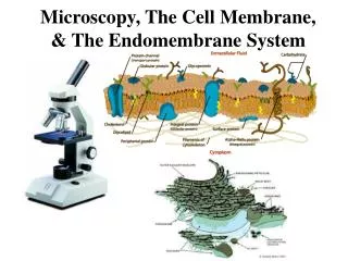
Microscopy, The Cell Membrane, & The Endomembrane System
140 likes | 514 Views
Microscopy, The Cell Membrane, & The Endomembrane System. Units of Cell Size. Virus .02 – 0.25 um (20 – 250 nm). Prokaryotic cell: 2 – 10 um. Eukaryotic cell: 10 – 100 um. Types of Microscopes: Compound Light Microscopes vs. Electron Microscopes . Electron Microscopes: SEM TEM.

Microscopy, The Cell Membrane, & The Endomembrane System
E N D
Presentation Transcript
Units of Cell Size Virus .02 – 0.25 um (20 – 250 nm) Prokaryotic cell: 2 – 10 um Eukaryotic cell: 10 – 100 um
Types of Microscopes: Compound Light Microscopes vs. Electron Microscopes
Electron Microscopes: SEM TEM
Calculating Total Magnification Total magnification = Magnification of Eyepiece X Magnification of Objective Lens
Calculating Linear Magnification Magnification = Size of the Image Actual Size of the Object Actual object size = Size of the Image Magnification The image size of this E.coli cell is 43 mm, and its actual size is 2um. Using this information, calculate the magnification of the image.
Making a Wet Mount Slide With your group… Create a wet mount slide of onion epidermal cells Sketch what your cells look like under the microscope (make sure to include the magnification on your sketch) Create a wet mount slide using salt water. Let your slide sit for 30 minutes before sketching and making observations!
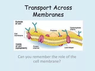
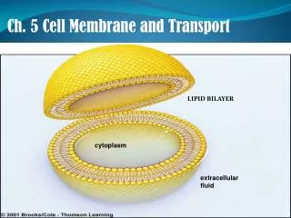
The Cell Membrane Review: The Fluid Mosaic Model A B F C E D
Endocytosis and Exocytosis: What other organelles are involved?
Endocytosis: A Closer Look Phagocytosis Pinocytosis (Cell Eating) (Cell Drinking) LIQUID! NON-SPECIFIC! SOLID MATERIAL! RECEPTOR MEDIATED! Amoeba phagocytosis Macrophage (white blood cell) phagocytosis Liver cells