Tomography
This article examines the limitations of planar X-ray angiography, particularly its inability to provide detailed 3D structural information. The text discusses how additional views enhance image quality, and how distance-weighted back projection can improve reconstruction in X-ray imaging. It also covers computed tomography (CT) and magnetic resonance imaging (MRI), highlighting the contrast mechanisms, such as T1 weighting and the importance of metabolic markers in PET scans. Additionally, it explores advances in MicroCT and the implementation of more accurate reconstruction algorithms.


Tomography
E N D
Presentation Transcript
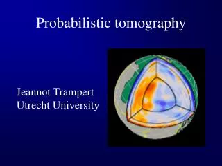
Limitation of Planar X-Rays Angiography as one solution (Kandel et al. Principles of Neural Science)
Tomography • If all we have is a single view (a) the information we have about 3-D structure can be represented by back-projecting those intensities along the direction of view • If we add more directions, the quality of the image improves (g = 160 views) (Russ, Image Processing Handbook.)
Weighted back projection • In the case of X-rays, there is a shadowing effect: if all the X-rays are absorbed by a structure close to the source, there is no contrast for deeper-lying structures • Solution: distance-weighted back projection: structures closer to the source are weighted more heavily in the reconstruction
Brain CT Wikipedia, Computed Axial Tomography
Other tomographic techniques - MRI • Nuclear dipole moment – aligns parallel to applied magnetic field • RF pulses applied perpendicular to magnetic field: knock dipole movement off • As nuclei process back to parallel, they emit radio waves • T1 weighting: time for nuclei to realign
T1 weighting Less dense tissue (CSF) relaxes fast = dark) Denser tissue (bone, myelin) relaxes more slowly (white) Wikipedia, Magnetic Resonance Imaging
PET scanning • Positron emission and positron annihilation • Deoxyglucose as a metabolic marker • 18F Deoxyglucose as a metabolic positron emitter
PET scan of brain Wikipedia, Positron Emission Tomography
MicroCT • Object is placed in a conical beam of X-rays • The closer the object is to the X-ray source, the greater the magnification • SkyScan 1072: 5 micron resolution
Fan beam reconstruction • X-ray beams through specimen are not parallel • Fan beam CT restricts X-rays to a fan • Reconstruction back-projects along fan of beams • Algorithm is fast, but not optimal
Fan Beam reconstruction Rosenfeld and Kak, Digital Picture Processing
Feldkamp cone beam reconstruction • X-ray beam is not constrained to a fan • Reconstruction is done for a cone of projections through the specimen • More accurate than fan-beam, but much slower • Newer algorithms use wavelet or Fourier transforms, rather than weighted back-projection
MicroCT of Rat Tibia Skyscan Web Site (www.skyscan.be)