Download

1 / 64
730 likes | 1.24k Views
Cell Signaling and Endocrine Regulation. Myxobacteria (soil-dwelling bacteria). Dictyostelium discoideum (cellular slime mold). Vibrio fischeri (marine bioluminiscent bacterium).

E N D
Vibrio fischeri (marine bioluminiscent bacterium) The Hawaiian bobtail squid (Eupryma scolopes) has a unique mutually beneficial (symbiotic) relationship with the bioluminescent bacterium Vibrio fischeri. (Image from M. J. McFall-Ngai and E. G. Ruby, University of Hawaii; National Science Foundation) Free-living Vibrio fischeri
Pheromonal communication in insects Tandem running of Procornitermes araujoi Emerson. • the male follows the large female, which acts as a leader in this process. The honey bee queen produces pheromones that help her: • attracting a retinue of workers around her, • attracting drones on mating flights, • preventing workers from reproducing at the individual (worker egg laying) and colony (swarming) level • regulating several other aspects of colony functioning.
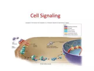
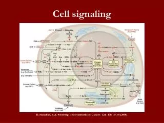
Cellular Communication Signalling Cell Target Cell signal (Chemical messenger) Response • Everything an animal does involves communication among cells • Example: moving, digesting food • Cell signaling – communication between cells
Cell Signalling Indirect Direct Long Distance Short Distance
Gap Junction Figure 3.2
Indirect Signaling Table 3.1
Glands Figure 3.3
Chemical Messengers Chemical messengers Hydrophobic/ Lypophilic Hydrophilic Other Lipids Peptides Purines Amines Steroids Gases • Structure of chemical messenger (especially hydrophilic vs. hydrophobic) affects signaling mechanism
Indirect Signaling Table 3.2
Peptide Hormones - Synthesis & Secretion Figure 3.4
Synthesis & Secretion of AVP/ADH Figure 3.5
Botulinum toxin • a protein produced by the bacterium Clostridium botulinum • affects the regulated exocytosis of neurotransmitters, preventing muscles contraction.
Transmembrane Receptor Figure 3.6
Steroid Hormones Smooth ER Cholesterol Mitochondria Steroids Reproductive hormones Glucocorticoides Mineralocorticoids Electrolyte balance Stress hormones Sex-specific characteristics
Steroid Hormones • Hydrophobic • Can diffuse through plasma membrane • Cannot be stored in the cell • Must be synthesized on demand • Transported to target cell by carrier proteins • Example: albumin • Bind to intracellular or transmembrane receptors • Slow effects on target cell (gene transcription) • Stress hormone cortisol has rapid non-genomic effects
Steroid Hormones Figure 3.8
Amine Hormones/ Biogenic Amines • Chemicals that possess amine group (–NH2) • Example: acetylcholine, catecholamines (dopamine, norepinephrine, epinephrine), serotonin, melatonin, histamine, thyroid hormones • Some true hormones, some neurotransmitters, some both • Most hydrophilic • Thyroid hormones are hydrophobic • Diverse effects
Other Chemical Messengers • Eicosanoids • Most act as paracrines • Hydrophobic • Often involved in inflammation and pain • Example: prostaglandins, leukotrienes • Gases • Most act as paracrines • Example: nitric oxide (NO), carbon monoxide • Purines • Function as neuromodulators and paracrines • Example: adenosine, AMP, ATP, GTP Figure 3.10
Communication to the Target Cell • Receptors on target cell • Hydrophilic messengers bind to transmembrane receptor • Hydrophobic messengers bind to intracellular receptors • Ligand • Chemical messenger that can bind to a specific receptor • Receptor changes shape when ligand binds
Ligand-Receptor Interactions • Ligand-receptor interactions are specific • Only the correctly shaped ligand (natural ligand) can bind to the receptor • Ligand mimics • Agonists – activate receptors • Antagonists – block receptors • Many ligand mimics act as drugs or poisons
Ligand-Receptor Interactions • A ligand may bind to more than one type of receptor • Receptor isoforms • Expressed on different target cells • Different responses to the same ligand • A single cell may have receptors for many different ligands
Ligand-Receptor Binding Figure 3.12
Changes in Number of Receptors • Number of receptors affects number of L-R complexes • More receptors L-R complexes response • Target cells can alter receptor number • Down-regulation • Target cell decreases the number of receptors • Often due to high concentration ligand • Up-regulation • Target cell increases the number of receptors
Changes in Number of Receptors Figure 3.13a
Ligand-Receptor Dynamics • Affinity of receptor for ligand affects number of L-R complexes • Higher affinity constant (Ka) response Figure 3.13b
Inactivation of Ligand-Receptor Complex • L-R complex must be inactivated to allow responses to changing conditions Figure 3.14
Signal Transduction Pathways • Convert the change in receptor shape to an intracellular response • Four components • Receiver • Ligand binding region of receptor • Transducer • Conformational change of the receptor • Amplifier • Increase number of molecules affected by signal • Responder • Molecular functions that change in response to signal
Transduction Pathway Figure 3.15
Types of Receptors Figure 3.16
Intracellular Receptors Figure 3.17
Changes in Gene Transcription Figure 3.18
Ligand-Gated Ion Channels Figure 3.19
Receptor Enzymes Figure 3.20
G-Protein-Coupled Receptors Figure 3.25
Second Messengers Table 3.3
Inositol-Phospholipid Signaling Figure 3.26
Cyclic-AMP Signaling Figure 3.27
Interaction Among Transduction Pathways • Cells have receptors for different ligands • Different ligands activate different transduction pathways • Response of the cell depends upon the complex interaction of signaling pathways
Regulation of Cell Signaling Regulated variable Biological Control Systems Response Integrating center Signal Signal Sensor Effector • Cell signaling is important for regulation of physiological processes • Components of biological control systems:
Regulation of Cell Signaling • Set Point • The value of the variable that the body is trying to maintain • Feedback loops • Positive • Output of effector amplifies variable away from the set point • Positive feedback loops are not common in physiological systems • Negative • Output of effector brings variable back to the set point
Feedback Regulation Figure 3.28
Pituitary Hormones • Pituitary gland secretes many hormones • Two distinct anatomic sections: • Anterior pituitary (adenohypophysis) • Posterior pituitary (neurohypophysis)
Posterior Pituitary • Extension of the hypothalamus • Neurons that originate in hypothalamus terminate in posterior pituitary • Neurohormones oxytocin and vasopressin synthesized in cell body and travel in vesicles down axons • First-order endocrine pathway • Hypothalamus receives sensory input • Hypothalamus serves as integrating center
Posterior Pituitary Figure 3.29
Anterior Pituitary • Hypothalamus synthesizes and secretes neurohormones • • Hypothalamic-pituitary portal system • • Anterior pituitary releases hormones • Tropic hormones • Cause release of another hormone • Third-order endocrine pathway
Anterior Pituitary Figure 3.30
Hypothalamus and Anterior Pituitary Figure 3.31