Download

1 / 54
540 likes | 647 Views
Homeostasis and Endocrine Signaling. 0. 32. What is osmosis?. Why is it important in cells?. Concept 32.3: A shared system mediates osmoregulation and excretion in many animals.

E N D
What is osmosis? Why is it important in cells?
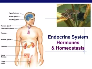
Concept 32.3: A shared system mediates osmoregulation and excretion in many animals Osmoregulation is the general term for the processes by which animals control solute concentrations in the interstitial fluid and balance water gain and loss
Osmosis and Osmolarity Cells require a balance between uptake and loss of water Osmolarity, the solute concentration of a solution, determines the movement of water across a selectively permeable membrane If two solutions are isoosmotic, the movement of water is equal in both directions If two solutions differ in osmolarity, the net flow of water is from the hypoosmotic to the hyperosmotic solution
Figure 32.15 (a) Osmoregulation in a marine fish Excretion of salt ions from gills Gain of water and salt ions from food Osmotic water loss through gills and other parts of body surface SALT WATER Gain of water and salt ions from drinking seawater Excretion of salt ions and small amounts of water in scanty urine from kidneys Key Water (b) Osmoregulation in a freshwater fish Salt Uptake of salt ions by gills Gain of water and some ions in food Osmotic water gain through gills and other parts of body surface Excretion of salt ions and large amounts of water in dilute urine from kidneys FRESH WATER
Marine and freshwater organisms have opposite challenges Marine fish drink large amounts of seawater to balance water loss and excrete salt through their gills and kidneys Freshwater fish drink almost no water and replenish salts through eating; some also replenish salts by uptake across the gills
What do you think the difference between osmoregulators and osmoconformers might be?
Osmoregulatory Challenges and Mechanisms Osmoconformers, consisting of some marine animals, are isoosmotic with their surroundings and do not regulate their osmolarity Osmoregulators expend energy to control water uptake and loss in a hyperosmotic or hypoosmotic environment
What are the 3 different ways you can excrete nitrogenous wastes?
Figure 32.16 Proteins Nucleic acids Amino acids Nitrogenous bases Amino groups Most aquatic animals, including most bony fishes Mammals, most amphibians, sharks, some bony fishes Many reptiles (including birds), insects, land snails Urea Uric acid Ammonia
Nitrogenous Wastes The type and quantity of an animal’s waste products may greatly affect its water balance Among the most significant wastes are nitrogenous breakdown products of proteins and nucleic acids Some animals convert toxic ammonia (NH3) to less toxic compounds prior to excretion
Ammonia excretion is most common in aquatic organisms Vertebrates excrete urea, a conversion product of ammonia, which is much less toxic Insects, land snails, and many reptiles including birds excrete uric acid as a semisolid paste It is less toxic than ammonia and generates very little water loss, but it is energetically more expensive to produce than urea
Figure 32.18 Nucleus of cap cell Cilia Flame bulb Interstitial fluid filters through membrane. Tubule Opening in body wall Tubules of protonephridia Tubule cell
Invertebrates Flatworms have excretory systems called protonephridia,networks of dead-end tubules connected to external openings The smallest branches of the network are capped by a cellular unit called a flame bulb These tubules excrete a dilute fluid and function in osmoregulation
In insects and other terrestrial arthropods, Malpighian tubules remove nitrogenous wastes from hemolymph without a filtration step Insects produce a relatively dry waste matter, mainly uric acid
Vertebrates In vertebrates and some other chordates, the kidney functions in both osmoregulation and excretion The kidney consists of tubules arranged in an organized array and in contact with a network of capillaries The excretory system includes ducts and other structures that carry urine from the tubules out of the body Animation: Nephron Introduction
Figure 32.19a Excretory Organs Posterior vena cava Kidney Renal artery and vein Aorta Ureter Urinary bladder Urethra
Figure 32.19b Kidney Structure Renal cortex Renal medulla Nephron Organization Renal artery Renal vein Afferent arteriole from renal artery Glomerulus Bowman’s capsule Ureter Proximal tubule Renal pelvis Peritubular capillaries Distal tubule Nephron Types Cortical nephron Efferent arteriole from glomerulus Branch of renal vein Descending limb Renal cortex Loop of Henle Vasa recta Collecting duct Ascending limb Renal medulla Juxtamedullary nephron
Many animal species produce urine by refining a filtrate derived from body fluids Key functions of most excretory systems Filtration: Filtering of body fluids Reabsorption: Reclaiming valuable solutes Secretion: Adding nonessential solutes and wastes from the body fluids to the filtrate Excretion: Releasing processed filtrate containing nitrogenous wastes from the body
Figure 32.17 Filtration Capillary Filtrate Excretory tubule Reabsorption Secretion Urine Excretion
Concept 32.4: Hormonal circuits link kidney function, water balance, and blood pressure The capillaries and specialized cells of Bowman’s capsule are permeable to water and small solutes but not blood cells or large molecules The filtrate produced there contains salts, glucose, amino acids, vitamins, nitrogenous wastes, and other small molecules
From Blood Filtrate to Urine: A Closer Look Proximal tubule Reabsorption of ions, water, and nutrients takes place in the proximal tubule Molecules are transported actively and passively from the filtrate into the interstitial fluid and then capillaries Some toxic materials are actively secreted into the filtrate
Descending limb of the loop of Henle Reabsorption of water continues through channels formed by aquaporin proteins Movement is driven by the high osmolarity of the interstitial fluid, which is hyperosmotic to the filtrate The filtrate becomes increasingly concentrated all along its journey down the descending limb
Ascending limb of the loop of Henle The ascending limb has a transport epithelium that lacks water channels Here, salt but not water is able to move from the tubule into the interstitial fluid The filtrate becomes increasingly dilute as it moves up to the cortex
Distal tubule The distal tubule regulates the K+ and NaCl concentrations of body fluids The controlled movement of ions contributes to pH regulation
Collecting duct The collecting duct carries filtrate through the medulla to the renal pelvis Most of the water and nearly all sugars, amino acids, vitamins, and other nutrients are reabsorbed into the blood Urine is hyperosmotic to body fluids Animation: Bowman’s Capsule Animation: Collecting Duct Animation: Loop of Henle
Figure 32.20 4 5 3 2 1 3 Proximal tubule Distal tubule NaCI Nutrients H2O H2O HCO3− HCO3− K NaCI NH3 H H K Interstitial fluid CORTEX Thick segment of ascending limb Descending limb of loop of Henle Filtrate H2O NaCI Salts (NaCI and others) H2O HCO3− OUTER MEDULLA NaCI H Urea Thin segment of ascending limb Collecting duct Glucose, amino acids Some drugs Urea NaCI H2O Key INNER MEDULLA Active transport Passive transport
Figure 32.20a 1 4 Proximal tubule Distal tubule Nutrients NaCI H2O H2O HCO3− K HCO3− NaCI NH3 H H K Filtrate Interstitial fluid CORTEX Active transport Passive transport
Figure 32.20b 3 2 3 5 Thick segment of ascending limb Descending limb of loop of Henle NaCI H2O OUTER MEDULLA NaCI Thin segment of ascending limb Collecting duct Active transport Passive transport Urea NaCI H2O INNER MEDULLA
Concentrating Urine in the Mammalian Kidney The mammalian kidney’s ability to conserve water is a key terrestrial adaptation
Figure 32.21-1 300 mOsm/L 300 300 300 H2O CORTEX 400 400 H2O Active transport Passive transport H2O H2O OUTER MEDULLA 600 600 H2O H2O 900 900 H2O INNER MEDULLA 1,200 1,200
Figure 32.21-2 300 mOsm/L 300 100 300 100 300 H2O NaCI CORTEX 400 200 400 NaCI H2O Active transport Passive transport H2O NaCI H2O NaCI OUTER MEDULLA 400 600 600 H2O NaCI H2O NaCI 900 700 900 NaCI H2O INNER MEDULLA 1,200 1,200
Figure 32.21-3 300 mOsm/L 300 100 300 100 300 300 H2O NaCI H2O CORTEX 400 200 400 400 NaCI H2O H2O Active transport NaCI Passive transport H2O H2O NaCI NaCI H2O H2O NaCI OUTER MEDULLA 400 600 600 600 H2O H2O NaCI Urea H2O NaCI H2O 900 700 900 Urea NaCI H2O H2O INNER MEDULLA Urea 1,200 1,200 1,200
Water and salt are reabsorbed from the filtrate passing from Bowman’s capsule to the proximal tubule In the proximal tubule, filtrate volume decreases, but its osmolarity remains the same As filtrate flows down the descending limb of the loop of Henle, water leaves the tubule, increasing osmolarity of the filtrate Salt diffusing from the ascending limb maintains a high osmolarity in the interstitial fluid of the renal medulla
The loop of Henle and surrounding capillaries act as a type of countercurrent system This system involves active transport and thus an expenditure of energy Such a system is called a countercurrent multiplier system
The filtrate in the ascending limb of the loop of Henle is hypoosmotic to the body fluids by the time it reaches the distal tubule The filtrate descends to the collecting duct, which is permeable to water but not to salt Osmosis extracts water from the filtrate to concentrate salts, urea, and other solutes in the filtrate
Adaptations of the Vertebrate Kidney to Diverse Environments Variations in nephron structure and function equip the kidneys of different vertebrates for osmoregulation in their various habitats
Desert-dwelling mammals excrete the most hyperosmotic urine and have long loops of Henle Birds have shorter loops of Henle but conserve water by excreting uric acid instead of urea
Mammals control the volume and osmolarity of urine The kidneys of the South American vampire bat can produce either very dilute or very concentrated urine This allows the bats to reduce their body weight rapidly or digest large amounts of protein while conserving water
Homeostatic Regulation of the Kidney A combination of nervous and hormonal inputs regulates the osmoregulatory function of the kidney These inputs contribute to homeostasis for blood pressure and volume through their effect on amount and osmoregulatory of urine
Antidiuretic Hormone Antidiuretic hormone (ADH) makes the collecting duct epithelium temporarily more permeable to water An increase in blood osmolarity above a set point triggers the release of ADH, which helps to conserve water Decreased osmolarity causes a drop in ADH secretion and a corresponding decrease in permeability of collecting ducts Animation: Effect of ADH
Figure 32.23-1 Osmoreceptors trigger release of ADH. ADH STIMULUS: Increase in blood osmolarity
Figure 32.23-2 Osmoreceptors trigger release of ADH. Thirst ADH Increased permeability Distal tubule STIMULUS: Increase in blood osmolarity Collecting duct
Figure 32.23-3 Osmoreceptors trigger release of ADH. Thirst Drinking of fluids ADH Increased permeability Distal tubule STIMULUS: Increase in blood osmolarity H2O reabsorption Collecting duct Homeostasis
The Renin-Angiotensin-Aldosterone System The renin-angiotensin-aldosterone system (RAAS) also regulates kidney function A drop in blood pressure near the glomerulus causes the juxtaglomerular apparatus (JGA) to release the enzyme renin Renin triggers the formation of the peptide angiotensin II
Figure 32.24-1 Distal tubule JGA releases renin. Renin Juxtaglomerular apparatus (JGA) STIMULUS: Low blood pressure
Figure 32.24-2 Liver Distal tubule Angiotensinogen JGA releases renin. Renin Juxtaglomerular apparatus (JGA) Angiotensin I ACE Angiotensin II STIMULUS: Low blood pressure