Download
1 / 41
480 likes | 848 Views
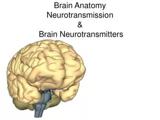
Introduction to brain anatomy. The brain. The brain in FSLview. sagittal. coronal. ACPC. axial. Terminology. Dorsal/superior. Right?. Rostral /anterior. Left?. caudal/posterior. Ventral/inferior. Superior/Dorsal surface. Inferior/Ventral surface. Anterior. Anterior. Rostral.

E N D
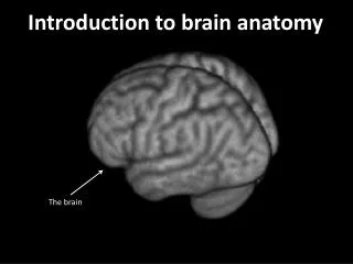
Introduction to brain anatomy The brain
The brain in FSLview sagittal coronal ACPC axial
Terminology Dorsal/superior Right? Rostral/anterior Left? caudal/posterior Ventral/inferior
Superior/Dorsal surface Inferior/Ventral surface Anterior Anterior Rostral Rostral Left Right Right Left Posterior Posterior Caudal Caudal
Grey Matter White Matter Constituent Tissues The brain is full of neurons. These are organised into two types of “tissues”: - Grey Matter - White Matter Post-Mortem MRI Neurons
Cerebral cortex Cerebral cortex • Makes up the bulk of the brain in humans • Newest part of the brain (in evolutionary terms) • Does thinking • Also most adaptable and variable part of brain
Two major modulatory systems cerebellum basal ganglia
Principle of organization: The cortex has sub-regions with different functions
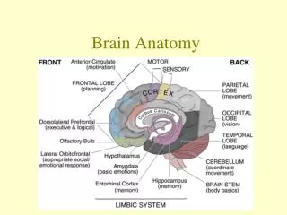
The cortex can be divided into 4 lobes Central sulcus frontal . parietal frontal parietal occipital occipital temporal Sylvian fissure Lateral surface Medial surface You should memorize these!
The cortex can be divided into 4 lobes Central sulcus parietal frontal parietal frontal occipital occipital temporal Sylvian fissure Lateral surface Medial surface You should memorize these!
Brodmann’s areas – (1909) • Divides cortex into 52 areas • Based on cytoarchitecture (which types of cells are present?) • Largely symmetrical (across two cerebral hemispheres) Don’t try to memorize these!
Modern cytoarchitectonics – Jülich atlas • Based on 10 brains • Registered into MNI space (affine) • Available in FSLview (atlas tools) • Disadvantage – subjects have to be dead
Gross anatomical features (sulci and gyri) e.g. Harvard-Oxford atlas in FSLview Problem – gyri do not correspond to functional regions Even if we could work out correspondence in one person, gyrification differs between individuals
Principle of organization: Function and connectivity are linked
Function and connectivity are linked • SMA and pre SMA • No obvious anatomical boundary • Different functional regions (top row) – for finger tapping and counting backwards in 3’s • Connectivity (DTI) based parcellation (bottom row) • Structure and function same dividing line between SMA and pre SMA Johansen-Berg et al (2004) PNAS 101(36):13335-40
Naming brain regions • A number of different systems are in use, most are arcane • Many areas will have a number of roughly-corresponding names • Brodmann areas (but only some of these are in common use) • Descriptive anatomical names e.g. dlPFC • Decoding: dl PF C • … but beware, some of these anatomical descriptions relate to the monkey brain!!! • 3. Descriptive names (often in Latin, e.g. cingulate) • 4. Functional names, e.g. visual cortex prefrontal cortex d=dorsal, v=ventral l=lateral, m=medial
Humans ≠ monkeys Monkey brain areas may have homologues in the human brain Not quite that simple…
Principle of organization: The brain contains maps of the outside world
The brain contains maps of the outside world 1. Somatotopy Sereno et al 1995 • Size of representation proportional to sensory/motor acuity • Adjacent parts of body are generally adjacent
The brain contains maps of the outside world 2. Retinotopy Dougherty et al (2003), Journal of Vision 3(10):586-598
What about the sub-cortical brain structures? Some software only shows the cortex Freesurfer Caret Advantage: can do cortical flattening, easier to compare cortical surface Disadvantage: gets rid of sub-cortical systems
Two major modulatory systems • Both interact heavily with cortex • Not just involved in motor system cerebellum basal ganglia
Basal ganglia Pharmacological diversity Many neurotransmitters and neuromodulators Imbalance linked to psychiatric disorders Two antagonistic pathways Direct and indirect Imbalance leads to disorders of movement and cognition Parkinson’s disease Huntington’s disease
Cerebellum Extremely regular micro circuitry Contains 50% of brain’s neurons Important for motor coordination but not only that
Principle of organization: Parallel circuits between cortex & subcortical structures
Subcortical-cortical loops 1. Thalamus Behrens et al (2003). Nat Neurosci. 6(7):750-7. • Correspondence between cortical regions and thalamic nuclei • They have reciprocal connections (thalamo-cortical and cortico-thalamic) • Thalamus also relays information from senses, basal ganglia and cerebellum to cortex
Subcortical-cortical loops 2. Basal ganglia Draganski et al (2008) J Neurosci. 28(28):7143-52
Subcortical-cortical loops 3. Cerebellum Dum and Strick (2003) J. Neurophysiology Lobules of the cerebellum connect to different cortical regions
Principle of organization: • Loops between cortex & subcortical structures • Each subcortical structure has a different contribution to information processing • This information processing function may be applied to many cortical areas • We can see many of the same principles of organization (functional localization, somatotopy) in subcortical structures • The corresponding bits of cortex & subcortical structures are interconnected in parallel & integrative loops
Top tips for finding your way around the brain
How to identify brain structures: 1. Use a brain atlas
How to identify brain structures: 2. Use the atlas toolbars in FSLview
How to identify brain structures: 3. Use a neuroscientist
Using a brain atlas These generally have axial, sagittal and coronal views Some structures are easier to identify in one view than another There are specialized atlases for some structures e.g. cerebellum and brainstem
Central sulcus Find the central sulcus in the axial view
The end! The brain