Download
1 / 41
570 likes | 1.2k Views
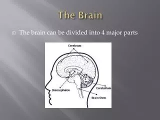
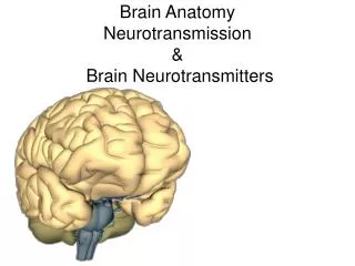
Brain Anatomy Neurotransmission & Brain Neurotransmitters. Brain Structures. Cerebral Hemisphere - 1/2 of Cerebrum. Cerebral Cortex. Cerebral Cortex (Grey Matter). The cerebrum’s surface—the cerebral cortex—is convoluted into hundreds of folds.

E N D
Cerebral Cortex Cerebral Cortex (Grey Matter) The cerebrum’s surface—the cerebral cortex—is convoluted into hundreds of folds. The cerebral cortex is where all the higher brain functions take place.
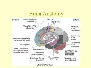
Cerebral Lobes • Frontal Lobe • Higher cognitive functions. • Problem solving • Memory • Parietal Lobe • Touch • Smell • Taste • Sensory and spatial awareness • Temporal Lobe • Emotions • Smelling • Tasting Occipital Lobe Vision Recognition Limbic Lobe
Limbic Lobe Cingulate cortex/ Limbic cortex The limbic lobe is located deep in the brain, and makes up the limbic system. The limbic system is the area of the brain that regulates emotion and memory. It directly connects the lower and higher brain functions Corpus callosum
The Basal Ganglia & Striatum Basal Ganglia/Striatum: Caudate nucleus Putament Deep below the cerebral cortex there are interconnected areas of grey matter collectively known as the "basal ganglia" (basement structures). Shown above (in green) is the largest structure in the basal ganglia called the striatum Play an important role in motivation, planning and coordinating motor movements and posture.
Brain Structures Brain Stem = (Midbrain+Hindbrain) - Cerebellum
Thalamus Thalamus means “inner room” in Greek, as it sits deep in the brain at the top of the brainstem. The thalamus is called the gateway to the cerebral cortex, as nearly all sensory inputs pass through it to the higher levels of the brain.
Hypothalamus • The hypothalamus sits under the thalamus at the top of the brainstem. Although the hypothalamus is small, it controls many critical bodily functions and set points: • Controls autonomic nervous system • Center for emotional response and behavior • Regulates body temperature • Regulates water balance and thirst • Controls sleep-wake cycles • Controls endocrine system The hypothalamus is shaded blue. The pituitary gland extends from the hypothalamus.
Brain Structures Brain Stem = (Midbrain+Hindbrain) - Cerebellum
The Brainstem • Most primitive part of the brain • controls the basic functions of life: breathing, heart rate, swallowing, reflexes to sight or sound, sweating, blood pressure, sleep, and balance. Midbrain Pons Medulla oblongata
Cerebellum The cerebellum is connected to the brainstem, and is the center for coordination of body movement and balance.
The Ventricles The ventricles are a complex series of spaces and tunnels through the center of the brain. The ventricles secrete cerebrospinal fluid (CSF), which suspends the brain in the skull. The ventricles also provide a route for chemical messengers that are widely distributed through the central nervous system.
Cerebrospinal Fluid Cerebrospinal fluid is a colorless liquid that bathes the brain and spine. It is formed within the ventricles of the brain, and it circulates throughout the central nervous system. Cerebrospinal fluid fills the ventricles and meninges, allowing the brain to “float” within the skull.
Blood-Brain Barrier The blood brain barrier consists of cells tightly packed around the capillaries of the CNS
Blood-Brain Barrier Drugs that cross the BBB and enter the brain must be non-protein bound, non-ionized, and highly lipid soluble
Brain Cells • Nerve Cells or Neuron • Variety of Supporting Cells • In CNS (brain and spinal cord) supporting cells consist mostly of neuroglial cells. neuron
Neurons & Neuron Structure Neurons are the basic building blocks of the nervous system. These specialized cells are the information-processing units of the brain responsible for receiving and transmitting information.
Signaling along Brain Circuits Signaling along brain circuits involves 2 steps • Nerve Impulse Conduction: • passage of an electrical signal (a positive deflection of the membrane voltage) along a neuron. • Neurotransmission or Synaptic Transmission: • The transmission of a nerve impulse from one neuron to another via a chemical signal.
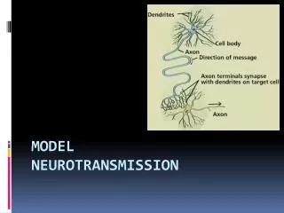
Neurotransmission (also called Synaptic Transmission) How nerve cells communicate with each other
Synaptic/Neurotransmission The process is quite complex but it is important to understand it because the synapse is a target for many common drugs The Synapse • Nervous impulses are transmitted from one neuron to another across structures called synapses. • Synapses separate neurons by a small gap called the synaptic cleft • Neurons communicate across the synaptic cleft using neurotransmitters. Post-synaptic terminal Pre-synaptic terminal
Steps in Neurotransmission • The nervous impulse (a +ve deflection of the voltage of the neuron) or action potential travels down the axon and arrives at the axon terminal. • The +ve deflection of the voltage of the axon terminal opens voltage-activated Calcium channels (pores in the membrane that are specifically permeable to Calcium ions) and Calcium ions enter the axon terminal. • Neurotransmitter molecules are present in the terminal of the neurons in little packages called synaptic vesicles.
Steps in Neurotransmission (contd) • In response to Calcium entry these synaptic vesicles fuse to the neuronal membrane and the neurotransmitters are released into the synaptic cleft. • Neurotransmitters bind to receptors on the post-synaptic membrane. • The activated post-synaptic receptors either stimulate (excite or depolarize, i.e make more +ve) or inhibit (hyperpolarize, i.e make more –ve) the postsynaptic neuron.
Termination of Neurotransmission • Neurotransmitter can diffuse away from the synapse. • Neurotransmitters may be broken down by enzymes present in the synapse. • Neurotransmitters may be pumped back into the presynaptic terminal (). • Once back in the presynaptic terminal they may be repackaged into synaptic vesicles. • Or they may be broken down by enzymes present in the presynaptic terminal (e.g. as in the case of monoamine neurotransmitters by the enzyme monoamine oxidase (MAO)).
1. Nerve Conduction How does a neuron or nerve fiber generate and conduct an electrical signal?
Resting Membrane Potentials & Basis for Neuronal Electric Currents The inside of a neuron is negatively charged with respect to the outside -60 mv
Intra- & Extra-Cellular Ionic Concentrations Concentration of Ions Inside a Neuron Concentration of Ions Outside a Neuron Intracellular K+ (140 mM) K+ (4 mM) Extracellular Na+ (4 mM) Na+ (140 mM) Cl- (4 mM) Cl- (140 mM) Ca++ (2 mM) Ca++ (0.1 nM) The Sodium (Na+)-Potassium (K+) Pump is responsible for maintaining High K+ and Low Na+ concentrations inside the neuron or cell. It is the target of many drugs.
K+ Channels are Open at Rest • The reason cells or neurons are negatively charged is that K+ (Potassium) channels are normally open at rest. • K+ will flow out of the neuron through these K+ specific pores (K+ channels) , taking away +ve charge and leaving the neuron with a net negative charge.
Ion Channels & Cellular Excitability • If a neuron or cell becomes more negatively charged it is said to be hyperpolarized and is less excitable or less likely to conduct a nerve impulse or action potential. • If a neuron or cell becomes more positively charged it is said to be depolarized and is more excitable or more likely to conduct a nerve impulse or action potential. -60 mV K+ (140 mM) K+ (4 mM) Na+ (4 mM) Na+ (140 mM) Cl- (4 mM) Cl- (140 mM) Ca++ (2 mM) Ca++ (0.1 nM)
Ion Channels & Cellular Excitability • Drugs that activate K+ channels make a neuron less excitable. • Drugs that activate Na+ channels ↑ excitability • Drugs that activate Cl- channels ↓ excitable • Drugs that activate Ca++ channels ↑ excitable -60 mV K+ (140 mM) K+ (4 mM) Na+ (4 mM) Na+ (140 mM) Cl- (4 mM) Cl- (140 mM) Ca++ (2 mM) Ca++ (0.1 nM)
Ion Channels & Cellular Excitability • K+ channels open→ hyperpolarization (↑ -ve), ↓ excitable • Na+ channels open→ depolarizaton (↑ +ve), ↑ excitable • Cl- channels open→ hyperpolarization (↑ -ve), ↓ excitable • Ca++ channels open→ depolarization (↑ +ve), ↑ excitable -60 mV K+ (140 mM) K+ (4 mM) Na+ (4 mM) Na+ (140 mM) Cl- (4 mM) Cl- (140 mM) Ca++ (2 mM) Ca++ (0.1 nM)
Action Potential • An action potential (or nerve impulse) is a transient alteration of the transmembrane voltage (or membrane potential) across an excitable membrane generated by the activity of voltage-gated ion channels embedded in the membrane (from Neuroscience. 4th ed.. Sinauer Associates). • Action potentials are pulse-like waves of +ve voltage that travel along axons of neurons and muscle cell including cardiac muscle.
Myelin and Propagation of Action Potentials • Many axons in the CNS are surrounded by oligodenrocytes that wrap around the axon forming a sheath of myelin. • Myelin provides electrical insulation that aids the propagation speed of the Action Potential. Nodes of Ranvier
Multiple Sclerosis Multiple sclerosis (MS), is an autoimmune disease of in which the body's immune response attacks a person's brain and spinal cord (CNS), leading to demyelination. Immune cells attacking myelin
How Neurotransmitters Generate Axon Potentials • Neurotransmitters either directly (by binding to ionotropic receptors) or indirectly (by binding to metabotropic receptors) activate ion channels expressed on the postsynaptic neuron. • These ion channels can either depolarize (excite or make more +ve) or hyperpolarize (inhibit or make more negative) the postsynaptic membrane. • If the sum of all the actions of the neurotransmitters acting at the postsynaptic membrane depolarizes (excites) the postsynaptic neuron sufficiently then the action potential chain reaction is initiated at the neuron.
Neurotransmitter Receptors Neurotransmitter Receptors are the targets of many drugs Ionotropic Receptors Are ion channels that open upon binding to neurotransmitter Neurotransmitter molecule Metabotropic Receptors Bind to neurotransmitters & modulate ion channels indirectly
Glutamate & GABA Neurotransmitters Glutamate • Primary excitatory neurotransmitter in the brain. • Major receptor is a non-specific cation channel GABA • Primary inhibitory neurotransmitter in the brain. • A major receptor is a Cl- ion channel.