Download
1 / 6

E N D
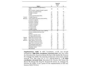
SupplementaryTable 1: ROIsCoordinates. L=Leftand R=righthemispheres. DMN ROIscoordinatesextractedfrom: [Fair DA, Cohen AL, Dosenbach NU, Church JA, Miezin FM, Barch DM, Raichle ME, Petersen SE, Schlaggar BL (2008). The maturingarchitectureofthebrain's default network. ProcNatlAcadSci U S A, 105(10):4028-32. ] CN ROIscoordinatesextractedfrom: [Dosenbach NU, Visscher KM, Palmer ED, Miezin FM, Wenger KK, Kang HC, Burgund ED, Grimes AL, Schlaggar BL, Petersen SE (2006). A core system for theimplementationoftask sets. Neuron, 50(5):799-812.]
Supplementary Figure 1: Head motiondescriptivestatistics - derivativeofthe RMS variance(DVARS) described in [Power JD, Barnes KA, Snyder AZ, Schlaggar BL, Petersen SE (2012). Spuriousbutsystematiccorrelations in functionalconnectivity MRI networks arisefromsubjectmotion. Neuroimage, 59(3):2142-54] and frame-wisedisplacement (FD) from [Jenkinson M, Bannister P, Brady M, Smith S (2002). Improvedoptimization for therobustandaccurate linear registrationandmotioncorrectionofbrainimages. Neuroimage, 17:825–841]
L. Parietal – R. Parahippo. Cerebellar Tons – aMPFC Left Parietal – aMPFC Left Parietal - Retrosplenial Post. Cingulate - vMPFC Retrosplenial – R.Parietal Left Parietal - vMPFC Left Frontal - aMPFC Right Parietal – aMPFC Retrosplenial – aMPFC Right Parietal – vMPFC Right Temporal - aMPFC R. Parahippo. – R. Frontal R. Parahippo. – aMPFC Supplementary Figure 2: Scatter-plot of the connectivity strength in the Default Mode Network with significant age effects. The labels of the brain region pairs are shown at the top of each panel. Each point represents a subject in the sample, and the red line depicts the predicted functional connectivity when a fitted linear model was considered. Adjusted-r and Bonferroni corrected p-values are highlighted in each panel. Abbreviation: aMPFC: anterior medial prefrontal cortex.
Degree: Left Parietal Degree: Posterior Cingulate Degree: Retrosplenial Degree: Right Temporal Degree: Right Parietal Degree: aMPFC EVC: Right Parahippocampal Degree: vMPFC EVC: Cerebellar Tons EVC: aMPFC Supplementary Figure 3: Scatter-plot of the graph descriptors’ value of the Default Mode Network with significant age effects. Each point on the graph represents the measure of a subject, and the red line depicts the predicted functional connectivity when the fitted linear model was considered. The Adjusted-r and Bonferroni corrected p-values are highlighted in each panel. Abbreviations: aMPFC: anterior medial prefrontalcortex; vMPFC: ventral medial prefrontalcortex; EVC: eigenvectorcentrality.
L. Anterior fusiform – R. Posterior Temporal L. Posterior Cingulate - RaIfO L. Posterior Cingulate - dACCmsFC R. Posterior Cingulate - dACCmsFC Supplementary Figure 4: Scatter-plot of the connectivity strength in the Control Network with significant age effects. The labels of the brain region pairs are shown at the top of each panel. Each point represents a subject of the sample, and the red line depicts the predicted functional connectivity when the fitted linear model was considered. Adjusted-r and Bonferroni corrected p-values are highlighted in each panel. Abbreviations: dACCmsFC: dorsal anterior cingulate/medial superior frontal cortex; aIfO: anterior insula/frontal operculum; EVC: eigenvectorcentrality.
Degree: Left Posterior Cingulate Degree: Left Posterior Temporal Degree: Right Posterior Cingulate Degree: RaIfO Degree: dACCmsFC Supplementary Figure 5: Scatter plot of the graph descriptors’ value of the Control Network with significant age effects. Each point on the graph represents a subject, and the red line depicts the predicted functional connectivity when the fitted linear model was considered. Adjusted-r and Bonferroni corrected p-values are highlighted in each panel. Abbreviations: dACCmsFC: dorsal anterior cingulate/medial superior frontal cortex; aIfO: anterior insula/frontal operculum; EVC: eigenvectorcentrality.