Objectives:
20 likes | 162 Views
USS Diagnosis of Gestational Trophoblastic Disease S Sivarajasingam, S Kar, J Chupi, S Visvanathan, N Deo Women and Children’s Services Whipps Cross University Hospital, Barts Health NHS Trust, London. Objectives:

Objectives:
E N D
Presentation Transcript
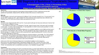
USS Diagnosis of Gestational Trophoblastic Disease S Sivarajasingam, S Kar, J Chupi, S Visvanathan, N Deo Women and Children’s Services Whipps Cross University Hospital, Barts Health NHS Trust, London Objectives: Our aim was to find the sensitivity of transvaginal ultrasonography (TVUS) in diagnosing women with gestational trophoblastic disease (GTD). We also looked at the outcome in these women. Methods: All histologically confirmed molar pregnancies at Whipps Cross University Hospital over a 10 year period, from January 2001 to February 2011, were audited. All of the confirmed molar pregnancies were referred to the Charing Cross GTD centre and follow up data was obtained from there. Results: 66 women were diagnosed with GTD over this 10 year period; although all notes were retrieved, 6 women had no scan reports available for review. The incidence of GTD was 1/714 deliveries. The age range of women was 19-45 years with an average of 30.9 years. 17 women were primigravidae, 27 were gravidae 2-4 while 5 women were gravidae 5 or more. In 17 women, exact parity was unknown. 28/66 women (42.4%) had a histological diagnosis of a complete hydatiform mole (CHM) and there was a pre-operative TVUS detection rate of 78.5%. The mean gestational age range at diagnosis was 7-24 weeks with an average of 11 weeks. 23 women (82%) presented with vaginal bleeding while 5 women (18%) were incidentally diagnosed at ultrasound. 36/66 women (54.5%) had a histological diagnosis of partial hydatiform mole (PHM). The TVUS detection rate was 27.8%. The gestational age range for detection was 5-21 weeks with an average of 13 weeks. 10 women (28%) presented with vaginal bleeding while 9 women (25%) were incidentally diagnosed at ultrasound. 17 women were excluded due to lack of documentation. 2/66 women (3%) had a histological diagnosis of unclassified hydatiform molar pregnancy. Both women who developed persistent trophoblastic disease had CHM. 3 women (5%) with a PHM inadvertently underwent medical management however PHM was suspected in one of these women by TVUS. It is noteworthy that no complications were seen in the medically managed PHM. Conclusion: GTD in a mixed ethnic background is still a rare diagnosis with an overall TVUS detection rate of 53.2% which is comparable to RCOG guidelines. The detection rate for both PHM and CHM were comparable to the ones quoted in recent literature which were 80% and 30% respectively. There has not been a significant change in diagnosis at an early gestational age however diagnosis in late pregnancy is rare, as demonstrated by this study with only 4/66 women diagnosed at a gestation greater than 18 weeks. (A) (B) References: Seckl et al, Lancet 2010 The Management of Gestational Trophoblastic disease, RCOG, February 2010 Alhamdan et al, Recognising gestational trophoblastic disease, Best Practice & Research Clinical Obstetrics & Gyanecology, 2009
USS diagnosis of Gestational Trophoblastic Disease • S Sivarajasingam, S Kar, J Chupi, D Visvanathan, N DeoWomen’s & Children’s Services • Whipps Cross University Hospital, Barts Health NHS Trust, London


