Download
1 / 57
570 likes | 840 Views
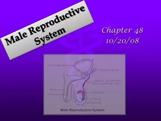
Male and Female Reproductive System video: Life's Greatest Miracle. MALE REPRODUCTIVE SYSTEM. Sperm – the male sex cell, can live up to 48 hours ,

E N D
Male and Female Reproductive Systemvideo: Life's Greatest Miracle
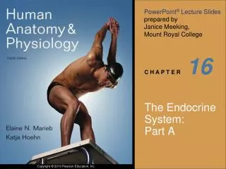
MALE REPRODUCTIVE SYSTEM Sperm – the male sex cell, can live up to 48 hours, A mature spermatozoa has 3 distinct parts: a head, a mid-piece, and a tail. The tail is made up of microtubules that form cilia and flagella, and the mid-piece contains energy-producing mitochondria. The head contains 23 chromosomes within a nucleus. The tip of the nucleus is covered by a cap called the acrosome, which is believed to contain enzymes needed to breach the egg for fertilization. 500 sperm end to end = 1 inch, 60 days for a sperm to mature, DNA is located in the head, tail used to travel 23 pairs of chromosomes, carries the sex cell X or Y, eggs only carry X, sperm determines sex of baby Humans have 46 chromosomes. Video ML part 1 6:47-9:04 1000 sperm per second, 100 million everyday Testes - where sperm is produced, Produces 100 million per day Epididymis– where sperm matures/stored in coiled tubes Vas Deferens – where sperm is stored and collected, 18 inches long Urethra- last tube the sperm exits through, passage way out of the body for semen and urine Ejaculatory duct – keeps urine and semen from mixing
HEAD TAIL MID SECTION
The penis has a long shaft and enlarged tip called the glans penis.. When the male is sexually aroused, the penis becomes erect and ready for intercourse. Erection is achieved because blood sinuses within the erectile tissue of the penis become filled with blood. The arteries of the penis are dilated while the veins are passively compressed so that blood flows into the erectile cartilage under pressure. The male penis is made of two different tissues, soft spongy tissue and cartilage.
Male baby Sex chromosomes
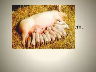
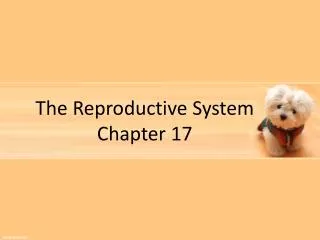
Bladder Seminal vesicle Ejaculatory duct Vas Deferens prostate Cowper’s gland urethra penis epididymis glans testes Scrotum
MALE Seminal Vesicle – dumps sucrose into the vas deferens, nutrients for sperm Cowper’s gland - dumps fluid that neutralizes urethra and vagina Prostate Gland – dumps fluid into urethra, urethra runs thru this gland Semen: all the fluid and sperm Why is the fluid important to the sperm? Helps it travel, gives nutrients and neutralizes the acidity in the female’s vagina and male’s urethra Video 4:56 Miracle of Life 2 Scrotum – sac which holds the testes, keeps sperm slightly below body temperature (98.6 degrees) The testes hang outside the abdominal cavity of the male within the scrotum. They begin their development in the abdominal cavity but descend into the scrotal sacs during the last 2 months of fetal development. This is required for the production of sperm because internal body temperatures are too high to produce viable sperm. Glans – tip of the penis
Foreskin: covering of skin that folds over the glans of the penis at birth Circumcision: surgical removal of the foreskin (ancient religions) Four years ago, a boy had part of his penis cut off during a circumcision. The part could have been reattached if his pediatrician had acted quickly enough to do so, according to the plaintiff. A Georgia jury agreed and awarded $1.8 million to the boy and $500,000 to his mother. Their names are being kept private, but the doctors' names aren't The hospital was not found to be at fault. The mother received $500,000 for psychotherapy, and the $1.8 million for the boy "was based on a per-day formula for the child's life expectancy."
David Reimer was born as a male identical twin in Winnipeg, Manitoba. His birth name was Bruce; his twin was named Brian. At the age of 6 months, after concern was raised about how they both urinated, the boys were diagnosed with phimosis. They were referred for circumcision at the age of 8 months. On April 27, 1966, a urologist performed the operation using the unconventional method of cauterization. The procedure did not go as doctors had planned, and Reimer's penis was burned beyond surgical repair • The parents, concerned about their son's prospects for future happiness and sexual function without a penis, took him to Johns Hopkins Hospital in Baltimore to see John Money, a psychologist who was developing a reputation as a pioneer in the field of sexual development and gender identity, based on his work with intersex patients. Money was a prominent proponent of the 'theory of Gender Neutrality'; that gender identity developed primarily as a result of social learning from early childhood and could be changed with the appropriate behavioral interventions. He and other physicians working with young children born with abnormal genitalia believed that a penis could not be replaced but that a functional vagina could be constructed surgically, and that he would be more likely to achieve successful, functional sexual maturation as a girl than as a boy.[4] • Brenda, estrogen….13 suicidal, 14 wanted to be male, brother dies overdose, 39 shoots himself
The bladder is emptied by way of the urethra, a tube passing through the prostate gland. The main function of the prostate is to supply fluid for sperm that has been collected in the seminal vesicles. Ejaculation is when the seminal vesicles and prostate empty.The seminal vesicles are supplied by the vas deferens from the epididymis, a tightly coiled, tube next to the testicle that provides for the storage, transmission, and maturation of sperm.Before ejaculation, the Cowper's glands secrete an alkaline fluid that neutralizes any urine that may be left in the urethra. The fluid also has a lubricating quality.
Pathway of the sperm Produces sperm testes dumps fluid Stored matures neutralizes epididymis v Prostate gland Cowper’s gland vas deferens Seminal vesicle transport exit from body dumps sucrose urethra
Cancer of the male Testicular cancer: self-exam page 482 less frequent than breast cancer, strikes young men between ages of 15-22. check for large lumps, or contour changes in shape etc. Testicles should feel smooth except raised organ located in the back. Prostate cancer: enlarged prostate can indicate risk of cancer, can lead to kidney and bladder trouble (age 50)
Medical News Today • Although testicular cancer is quite rare, it is the most common cancer in men between the ages of 20 and 45. But it can almost always be treated successfully. The testicles are located behind the penis in a sac called the scrotum. Testicular cancer may cause one or both of the testicles to enlarge or it may cause a lump in the scrotum. There may or may not be pain with the swelling. What you can do -- All men aged 15 or over should check their testicles regularly. -- Become familiar with your testicles so you can detect any changes early; report any changes to your doctor. -- Have regular medical check-ups by your doctor that include testicular examination. What to watch for -- Any change in size, shape, consistency, swelling or sensation of your testicles or scrotum -- Pain in the testicles or scrotum -- A dull ache or heaviness in your lower abdomen -- Unusual and persistent backache -- Unexplained weight loss
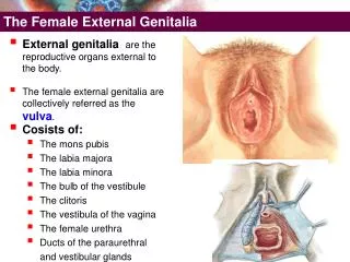
Female Reproductive System • Ova (ovum or egg) female sex cell of reproduction • ovary: where eggs (ova ) are produced size of ova .............................. 30 eggs All of the cells that will ever become eggs are present at birth. A baby girl holds approximately 1 ½ million ova in her ovaries. When she reaches 12 years old ,only a ¼ of these ova are alive. At puberty only 1 egg is released each month. • Ovulation: the ripening and release of an egg or ovum
A B D C
Menstruation There is a broad range of "normal" among menstruating women. The average cycle takes 28 days, but 21 to 35 days is considered normal for women in their 20s and 30s. Unpredictable or long menstrual cycles are normal for teenagers and women in their 40s. For teens, normal cycles can be as short as 21 days or as long as 45 days. If you are a teen, you can expect cycles to even out over time. If you are nearing the age of menopause, you can expect menstrual cycles to become longer and eventually to stop.
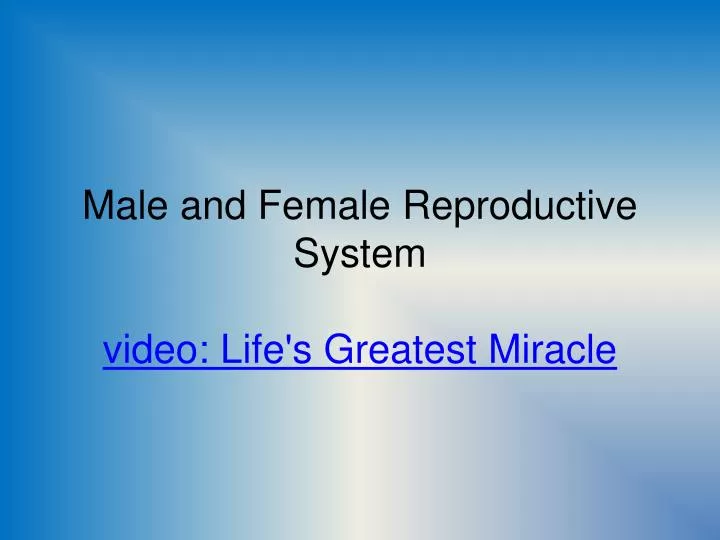
Fallopian tube: egg travels down this tube on the way to the uterus, fertilization takes place here • Cervix: opens into the vagina, dilates during birth • Uterus: also known as the womb… organ made of muscle which pushes the baby out. The uterus consists of a body and a cervix. The cervix protrudes into the vagina. • Vagina: last passage way out of the female • Video 1:40-3:30
…… Egg Ovary Uterus Fallopian tube Cervix Vagina
CONCEPTION • Every 28-30 days an egg ripens (matures) and is released from the ovary. It then travels to the fallopian tube where the egg can be fertilized by the sperm. • The fertilized egg or unfertilized egg then slowly travels to the uterus. Each month the uterus prepares far the fertilized egg. • Menstrual cycle: the cyclic ripening of an ovum and the preparation of the uterus for pregnancy. Also called ovulatory cycle • Fertilization: sperm and ova unite video ML3 all • Conception: the union of an ovum and sperm (implantation of the blastocyst into the wall of the mother’s uterus) LIFE? Contraceptives?
How does the uterus prepare for the fertilized egg? • The uterus lining builds up with soft tissue and rich blood supply. • About mid cycle(14 days)the ovum will burst from the ovary. (ovulation) • If the egg is fertilized it will embed itself into the lining of the uterus. • If the egg is not fertilized the egg and lining of the uterus is shed. (menstruation) • Menstruation lasts 4-5 days on average.
Pathway of the egg muscle ovary fallopian tubes uterus fertilization womb Immature eggs stored ovulation fertilized egg unfertilized egg menstruation egg implants in lining of uterus and begins to grow lining of uterus and egg is shed
Cancer of Female Reproductive System • Breast cancer: Breast cancer is a disease in which malignant (cancer) cells form in the tissues of the breast. self exam • One in eight women will be diagnosed with breast cancer in their lifetime. • Each year it is estimated that nearly 200,000 women will be diagnosed with breast cancer and more than 40,000 will die. Approximately 1,700 men will also be diagnosed with breast cancer and 450 will die each year
What is a mammogram? A mammogram is an x-ray picture of the breast. Mammograms can be used to check for breast cancer in women who have no signs or symptoms of the disease. This type of mammogram is called a screening mammogram. Screening mammograms usually involve two x-ray pictures, or images, of each breast. The x-ray images make it possible to detect tumors that cannot be felt. Screening mammograms can also find microcalcifications (tiny deposits of calcium) that sometimes indicate the presence of breast cancer.
Cervical Cancer • Causes and risk factors for cervical cancer have been identified and include human papillomavirus (HPV) infection, having many sexual partners, smoking, taking birth control pills, and engaging in early sexual contact. • HPV infection may cause cervical dysplasia, or abnormal growth of cervical cells. vaccines • Regular pelvic exams and Pap testing can detect precancerous changes in the cervix.
What is a Pap Test? The Pap test, also called a Pap smear, checks for changes in the cells of your cervix. The cervix is the lower part of the uterus (womb) that opens into the vagina (birth canal). The Pap test can tell if you have an infection, abnormal (unhealthy) cervical cells, or cervical cancer. Why do I need a Pap test? A Pap test can save your life. It can find the earliest signs of cervical cancer. If caught early, the chance of curing cervical cancer is very high. Pap tests also can find infections and abnormal cervical cells that can turn into cancer cells. Treatment can prevent most cases of cervical cancer from developing.
Beginning of LifeTeen pregnancy: There Goes My Life..Kenny Chesney • zygote: fertilized egg • Blastocyst: cluster of cells video ML4 • Embryo: cells after it implants (1st 8 weeks) • Fetus: 8 weeks until birth video ML5 • Pregnancy: 9 months or 280 days • Pregnancy test: blood or urine • Labor: contractions of the uterus
Amniotic sac: sac with fluid the surrounds embryo, acts as protector…shock absorber, regulates temperature…water breaking • Placenta: secures the embryo to the rich uterine lining…acts as lungs, digestive system, kidneys, protective barrier, and filtering system • Umbilical cord: carries blood back and forth from placenta to unborn child Contains blood vessels which flow between embryo and placenta • Miscarriage: spontaneous abortion , baby is born before it can survive • Caesarean: cutting into uterine wall to remove baby • Vaginal birth: baby is born thru the vagina
3 stages of Birth Stage 1 • Uterus contracts • Dilation of cervix • Longest stage • Stretching of cervix • Goal is 10 centimeters
Stage 2 • Cervix fully dilated • Uterus contracting • Head face down in birth canal Stage 3 • Expulsion of placenta • 10-15 minutes after birth
24-25 WEEKSA protective waxy substance called Vernix covers the skin. By birth, most of the Vernix will be gone but any that is left is quickly absorbed. Fetus has a hand and startle reflex. Footprints and fingerprints are forming. Fetus practices breathing by inhaling amniotic fluid into its developing lungs. At 24-25 weeks there is a 50-75% chance of survival if born, and is considered viable at this stage.
26-28WEEKSRapid brain development occurs during this period and the nervous system is able to control some bodily functions. The fetus’ eyelids now open and close. There is now a more than 90% chance of survival if the fetus is born at this point.
29-32 WEEKSThere is a rapid increase in the amount of body fat the fetus has. Rhythmic breathing occurs, but the lungs are not yet mature. The fetus sleeps 90-95% of the day. At this point the survival rate is above 95% if the baby is born.
38-40 WEEKSThe fetus is considered full-term. Lanugo is gone except on upper arms and shoulders. Hair on the baby’s head is now coarser and thicker. The lungs are mature. The average weight of the baby at this point is seven and a half pounds. At birth the placenta detaches from the uterus and the umbilical cord will be cut as the baby takes his first breaths of air. Breathing will trigger changes in the heart and bypass arteries forcing all blood to now travel through the lungs.