Brain & Cranial Nerves
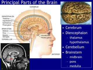
Brain & Cranial Nerves. The Brain. Ranges from 750 cc to 2100 cc Contains almost 98% of the body’s neural tissue Average weight about 1.4 kg (3 lb). 6 Major Regions of the Brain. Cerebrum Cerebellum Diencephalon Mesencephalon Pons Medulla oblongata. The Brain. Cerebrum.

Brain & Cranial Nerves
E N D
Presentation Transcript
The Brain • Ranges from 750 cc to 2100 cc • Contains almost 98% of the body’s neural tissue • Average weight about 1.4 kg (3 lb)
6 Major Regions of the Brain • Cerebrum • Cerebellum • Diencephalon • Mesencephalon • Pons • Medulla oblongata
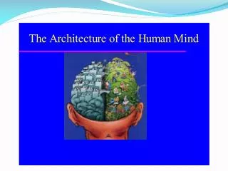
Cerebrum • Largest part of brain • Controls higher mental functions • Divided into left and right cerebral hemispheres • Surface layer of gray matter (neural cortex)
3 Functional Principles of the Cerebrum • 1. Each cerebral hemisphere receives sensory information from, and sends motor commands to, the opposite side of body
3 Functional Principles of the Cerebrum • 2. The 2 hemispheres have different functions although their structures are alike
3 Functional Principles of the Cerebrum • 3. Correspondence between a specific function and a specific region of cerebral cortex is not precise
Motor & Sensory Areas of the Cerebral Cortex • Where are the motor, sensory, and association areas of the cerebral cortex, and what are their functions?
Motor Areas • Precentral gyrus of frontal lobe: • directs voluntary movements • Primary motor cortex: • is the surface of precentral gyrus • Pyramidal cells: • are neurons of primary motor cortex
Sensory Area • Postcentral gyrus of parietal lobe: • receives somatic sensory information (touch, pressure, pain, vibration, taste, and temperature) • Primary sensory cortex: • surface of postcentral gyrus
Special Sensory Cortexes • Visual cortex: • information from sight receptors • Auditory cortex: • information from sound receptors • Olfactory cortex: • information from odor receptors • Gustatory cortex: • information from taste receptors
Sensory Association Areas • Somatic sensory association area: • interprets input to primary sensory cortex (e.g., recognizes and responds to touch) • Visual association area: • interprets activity in visual cortex • Auditory association area: • monitors auditory cortex
Other Integrative Areas • Speech center: • is associated with general interpretive area • coordinates all vocalization functions • Prefrontal cortex of frontal lobe: • integrates information from sensory association areas • performs abstract intellectual activities (e.g., predicting consequences of actions)
Hemispheric Lateralization • Functional differences between left and right hemispheres • Each cerebral hemisphere performs certain functions not performed by the opposite hemisphere
The Left Hemisphere • In most people, left brain (dominant hemisphere) controls: • reading, writing, and math • decision-making • speech and language
The Right Hemisphere • Right cerebral hemisphere relates to: • senses (touch, smell, sight, taste, feel) • recognition (faces, voice inflections)
Cerebellum • Second largest part of brain • Coordinates repetitive body movements • 2 hemispheres • Covered with cerebellar cortex
The Cerebellum • An autonomic processing center
Functions of the Cerebellum • Adjusts postural muscles • Fine-tunes conscious and subconscious movements
Structures of the Cerebellum • Folia: • surface of cerebellum • highly folded neural cortex • Anterior and posterior lobes: • separated by primary fissure
Structures of the Cerebellum • Cerebellar hemispheres: • separated at midline by vermis • Vermis: • narrow band of cortex • Flocculonodular lobe: • below fourth ventricle
Purkinje Cells • Large, branched cells • Found in cerebellar cortex • Receive input from up to 200,000 synapses
Arbor vitae • Highly branched, internal white matter of cerebellum • Cerebellar nuclei: • embedded in arbor vitae • relay information to Purkinje cells
The Peduncles • Tracts link cerebellum with brain stem, cerebrum, and spinal cord: • superior cerebellar peduncles • middle cerebellar peduncles • inferior cerebellar peduncles
Diencephalon • Composed of • Epithalmus • Hypothalamus • thalamus
The Thalamus • Final relay point for ascending sensory information • Coordinates the activities of the cerebral cortex
Hypothalamus • Controls somatic motor activities subconsciously • Rage, pleasure, pain, & sexual arousal • Controls autonomic function • Regulate heart rate, blood pressure, respiration, & digestive function • Coordinates activities of the nervous and endocrine systems • Inhibits or stimulates endocrine cells in the pituitary gland
Hypothalmus cont. • Secrete 2 hormones • Antidiuretic hormone – restricts water loss at the kidneys • Oxytocin –stimulates smooth muscle contractions • Uterus/mammary glands • Prostrate gland • Produce emotions & behavioral drives • Feeding center, thirst center
Hypothalmus cont. • Coordinate voluntary & autonomic functions • Think about a dangerous situation • Heart rate increases, respiratory rate goes up • Regulates body temperature • If temp. falls instructions sent to medulla oblongata to dialate blood vessels • Controls Circadian rhythms • 24 hour, day-night cycle • Retina of the eye
Medulla Oblongata • Connects brain and spinal cord
The Pons • Sensory & motor nuclei for 4 cranial nerves • Nuclei that help control respiration • Nuclei & tracts linking the cerebellum with the brain stem, cerebrum, and spinal cord • Ascending, descending & transverse tracts
Cranial Nerves • I – olfactory nerves – sensory • Sense of smell • II – optic nerves – sensory • Visual information • III – oculomotor – motor • Motor eye movements • IV – TrochlearNerves – motor • Superior oblique muscle
Cranial Nerves • V - Trigeminal Nerves - mixed, sensory • Motor – jaw, mastication, eyelid, eyebrow, lips, gums. teeth, cheek • Sensory – forehead, palate, nasal cavity • VI - Abducen Nerves - motor • Motor eye movement, lateral rectus • VII - Facial – mixed • Sensory – anterior 2/3 of tongue • Motor – lacrimal gland, nasal mucous gland, submandibular & sublingual salivary glands
Cranial Nerves • VIII – Vestibulocochlear Nerves - sensory • Special sensory – balance & equilibrium, hearing • IX – Glossopharyngeal – mixed • Sensory – posterior 1/3 of tongue, part of the pharynx & palate, carotid arteries • Motor – pharyngeal muscles, parotid salivary gland
Cranial Nerves • X – Vagus – mixed • Sensory – pharynx, diaphragm, • Motor – pharyngeal muscles, visceral organs in thorax & abdominal cavity • XI – Accessory Nerves - motor • Muscles of neck & upper back • XII – Hypoglossal Nerves – motor • Tongue movement