Download
1 / 66
690 likes | 1.2k Views
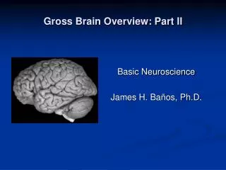
Gross Brain Overview: Part II . Basic Neuroscience James H. Baños, Ph.D. Overview. Organization Morphology Developmental/Evolutionary Cytoarchitectural 3-D Orientation to Internal Structures. How do we organize and characterize different parts of the brain?.

E N D
Gross Brain Overview: Part II Basic Neuroscience James H. Baños, Ph.D.
Overview • Organization • Morphology • Developmental/Evolutionary • Cytoarchitectural • 3-D Orientation to Internal Structures
How do we organize and characterize different parts of the brain?
How do we organize and characterize different parts of the brain? • Morphology • Developmental/Evolutionary Origins • Cytoarchitecture • Function
Frog Rat What’s changing? Cat Monkey Human
Sulci - The “valleys” on the surface of the brain • Gyri - The “Hills”
The term “gyrus” is sometimes used broadly and doesn’t always refer to a single well-defined ridge on the surface of the brain. The distinctions between large gyri are sometimes better seen in coronal sections.
Hemispheres Longitudinal Fissure
Lobes Central (Rolandic) Sulcus Lateral (Sylvian) fissure Parieto-occipital fissure
Frontal Parietal Temporal Occipital Lobes Cingulate Gyrus “Limbic Lobe”
Brain Stem Thalamus Hypothalamus Midbrain Pons Medulla
Cerebellum: Superior Aspect • 2 Hemispheres • Vermis Ant
Hemisphere Vermis Hemisphere Cerebellum: Posterior Aspect Anterior Lobe Primary Fissure Posterior Lobe
Cerebellum: Mid-Saggital Anterior Vermis Posterior Vermis
Developmental Origins • Areas of the Brain can be characterized by the embryonic origins of the tissue. • Nervous system begins as a tube that differentiates into three vessicles: • Prosencephalon • Mesencephalon • Rhombencephalon Three Vessicle Stage
Developmental Origins • Prosencephalon differentiates: • Telencephalon -- beginnings of hemispheres • Diencephalon • Rhombencehphalon differentiates • Metencephalon • Meyelencephalon • We use this terminology to describe the parts of the brain that develop from these vessicles Five Vessicle Stage
Organization • Telencephalon • Cortex • Basal Ganglia • Limbic System • Hippocampus
Organization • Diencephalon • Thalamus • Hypothalamus
Organization • Mesencephalon • Midbrain
Organization • Metencephalon • Cerebellum • Pons
Organization • Myelencephalon • Medulla
Major Internal Structures • Ventricular System • Amygdala (helpful landmark) • Thalamus/Hypothalamus/brain stem • Basal Ganglia • Caudate Nucleus • Putamen • Globus Pallidus • Hippocampal formation • Hippocampus • Fimbria • Fornix • Major white matter landmarks • Corpus callosum • Internal capsule
Evolution and Development Frog Rat Cat Monkey Human
Evolution and Development Frog Rat Cat Monkey Human ?
Evolution and Development Ventricles Basal Ganglia Hippocampus
Evolution and Development Why not the thalamus?
Ventricles Lateral Ventricles Third Ventricle Fourth Ventricle
Ventricles • Ventricles are connected (communicate) • Intraventricular Foramina (of Monroe) • Lateral Ventricles to Third Ventricle • Wide, oval hole • Cerebral Aqueduct (of Sylvius) • Third Ventricle to Fourth • Long, thin channel • Foramen of Magendie • Median aperture -- Fourth ventricle to subarachnoid space • Foramina of Luschka • Lateral apertures -- Fourth ventricle to subarachnoid space
Ventricles Foramen of Monroe Aqueduct of Sylvius Foramina of Luschka Foramen of Magendie
Choroid Plexus and CSF • Choroid Plexus • Spongy tissue located in the ventricles • Rich capillary bed • Pia Mater • Choroid endothelial cells • Produces CSF • About .35 ml per minute • Total volume 70-120 ml
CSF Flow • Lateral ventricles • Foramina of Monroe • 3rd ventricle • Aqueduct of Sylvius • 4th Ventricle • Foramen of Magendie/foramina of Lushka • Subarachnoid Space • Arachnoid granulations (absorption) • Superior sagittal sinus
CSF Absorption • CSF flows to the dorsal surface of the brain, where arachnoid granulations form a one-way valve and let the excess CSF enter the veinous drainage of the superior sagittal sinus