Download
1 / 3
30 likes | 187 Views
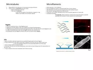
Microtubules. Microfilaments. Responsible for the separation of chromosomes during cell division. Grow out from a centrosome near the nucleus. R esist compression to the cell. Has a pair of centrioles , each has nine triplets of microtubules arranged in a ring.

E N D
Microtubules Microfilaments • Responsible for the separation of chromosomes during cell division. • Grow out from a centrosome near the nucleus. • Resist compression to the cell. • Has a pair of centrioles, • each has nine triplets of microtubules arranged in a ring. • Before a cell divides, the centrioles replicate. • Solid rods about 7 nm in diameter. • Each microfilament is built as a twisted double chain of actin subunits. • Microfilaments can form structural networks due to their ability to branch. • bear tension, resisting pulling forces within the cell. • important in cell motility, especially as part of the contractile apparatus of muscle cells. • amoeboid movement. • Pseudopodia, cellular extensions, extend and contract through the reversible assembly and contraction of actin subunits into microfilaments. • flagella. • There are usually just one or a few flagella per cell. • Flagella are the same width as cilia, but 10–200 microns long.eir beating patterns. • A flagellum has an undulatory movement that generates force in the same direction as the flagellum’s axis. • In spite of their differences, both cilia and flagella have the same ultrastructure. • The bending of cilia and flagella is driven by the arms of a motor protein, dynein. • cilia • Many unicellular eukaryotic organisms are propelled through water by cilia and flagella. • They can extend from cells within a tissue layer, beating to move fluid over the surface of the tissue. • Cilia in the windpipe sweep mucus carrying trapped debris out of the lungs. • Occur in large numbers on the cell surface. • Cilia move more like oars with alternating power and recovery strokes that generate force perpendicular to the cilium’s axis. • They are about 0.25 microns in diameter and 2–20 microns long.