Download
1 / 25
250 likes | 407 Views
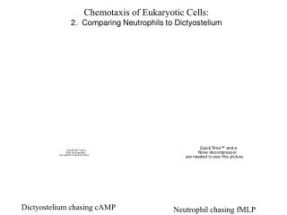
Chemotaxis of Eukaryotic Cells: Dictyostelium discoideum - Part 2 http://dictybase.org/Multimedia/index.html. http://dictybase.org/Multimedia/development/development.html. 2. Adjacent cells chemotaxis up the cAMP gradient and the phosphodiesterase they release degrades the cAMP.

E N D
Chemotaxis of Eukaryotic Cells: Dictyostelium discoideum - Part 2 http://dictybase.org/Multimedia/index.html http://dictybase.org/Multimedia/development/development.html
2. Adjacent cells chemotaxis up the cAMP gradient and the phosphodiesterase they release degrades the cAMP. 4. The cycle continues as the next set of cells chemotax toward the center of mass of cells and in turn release cAMP after a lag, recruiting yet more cells 3. After a lag, the chemotaxing cells release their own cAMP. This creates a a second, greater cAMP gradient. 1. At high density and low nutrient availability, a random cell begins to release cAMP.
cAMP release from a pipett stimulates chemotaxis of a population of Dictyostelium (from Peter Devreotes)
PIP2 PIP2 PH PH Recruitment of cortical actin to drive cell polarization and motility Adenylyl Cyclase cAMP (secreted) Outside cAMP Receptor PIP2 PIP2 PIP2 PIP2 PIP3 PIP3 PIP3 PIP3 ? ? PI3K ? ? Gbg Gbg PTEN Ga Ga Cytosol
A green fluorescent protein chimera of a pleckstrin homology (PH) domain that binds to the lipid phosphatidylinositol-3,4,5-trisphosphate (PIP3) reveals that this lipid is generated at the leading edge of the cell in a dynamic fashion that rapidly responds to changes in the extracellular cAMP gradient. (from Richard Firtel)
Iijima and Devreotes 2002 Cell 109, 599 GFP-PTEN localizes to the rear of a chemotaxing cell. The diamond indicates the location of the cAMP-containing pipette. A mutant of PTEN lacking the N-terminal 16 amino acids fails to localize to the rear - right panel.
Van Haastert and Devreotes 2004 Nat Rev Mol Cell Biol 5, 626 PI3K is not critical for acute stimulation of cortical actin accumulation but participates in remodeling of actin to from a polarized leading edge
Van Haastert and Devreotes 2004 Nat Rev Mol Cell Biol 5, 626
Van Haastert and Devreotes 2004 Nat Rev Mol Cell Biol 5, 626 ? ?
Local Excitation, Global Inhibition (LEGI) Model Ma et al., 2004 Biophysical J. 87, 3764 Assumptions: PI3K LEGI Receptor occupation rapidly stimulates a local, membrane imbedded (slowly diffusable) component that activates a membrane imbedded PI3K binding protein. This recruits PI3K from the cytosol to the membrane and results in local production of PIP3. Receptor occupation also stimulates a cytosolic component that diffuses throughout the cell and globally inactivates the membrane imbedded PI3K binding protein. The activation of the global inhibitor is slower than activation of the local activator. Thus, PI3K is concentrated near activated receptors
Local Excitation, Global Inhibition (LEGI) Model Ma et al., 2004 Biophysical J. 87, 3764 Assumptions: PTEN Regulation Receptor occupation rapidly stimulates a local, membrane imbedded (slowly diffusable) component that inactivates a membrane imbedded PTEN binding protein. This locally releases PTEN from the membrane and allows local production of PIP3. Receptor occupation also stimulates a cytosolic component that diffuses throughout the cell and globally activates the membrane imbedded PTEN binding protein. The activation of the global PTEN regulator is slower than activation of the local regulator. Thus, PTEN levels are reduced near activated receptors and elevated elsewhere. Note: PTEN regulation and PI3K regulation are assumed to be uncoupled
R PI3K half of the LEGI model L L S Membrane Membrane EA E Local BSAPI3K BSAPI3K BSAPI3K BSPI3K BSPI3K PI3K I IA PI3K PI3K IA Global Cytosolic PI3K, I and IA are freely diffusable throughout the cytosol. Membrane imbedded components (R, S, E, BS) have more restricted movement
R PTEN half of the LEGI model L L S Membrane Membrane EA E Local BSAPTEN BSAPTEN BSAPTEN BSPTEN BSPTEN PTEN IPTEN IAPTEN PTEN PTEN IAPTEN Global Cytosolic PTEN, I and IA are freely diffusable throughout the cytosol. Membrane imbedded components (R, S, E, BS) have more restricted movement
Virtual Cell Model Ma et al. (Devreotes & Iglesias) Using Lowe and Schaff, 2001, Trends Biotechnol. 19, 401; Virtual Cell http://www.nrcam.uchc.edu/applications/applications.html
Virtual Cell Model Ma et al. (Devreotes & Iglesias) IAPTEN creates PTEN binding sites IAPI3K eliminates PI3K binding sites
Virtual Cell Model Ma et al. (Devreotes & Iglesias) wt (2 fold) (10% wt for each)
Virtual Cell Model Ma et al. (Devreotes & Iglesias) PTEN cAMP PI3K PIP3
Virtual Cell Model Ma et al. (Devreotes & Iglesias) The PIP3 gradient depends on the cAMP gradient but is relatively insensitive to the absolute amount of cAMP, allowing adaptation to higher basal cAMP
Van Haastert and Devreotes 2004 Nat Rev Mol Cell Biol 5, 626 Myosin II accumulates in the lateral and trailing edge of the migrating cell. There it suppresses formation of additional pseudopods and retracts the trailing end of the cell. What is the mechanism for polarized location of myosin II and for activation of myosin II-dependent contraction?
Van Haastert and Devreotes 2004 Nat Rev Mol Cell Biol 5, 626 Elevation in cGMP stimulates the global formation of myosin II filaments and also activates myosin light chain kinase, which enhances traction on actin filaments. This drives retraction of pseudopods and retraction of the uropod tail. However, it does not explain why myosin II is excluded from the anterior region of the cell. Receptor Guanylate cyclase cGMP-binding protein Myosin light chain kinase
Van Haastert and Devreotes 2004 Nat Rev Mol Cell Biol 5, 626 cAMP cAR1 PI3K PIP3 AKT PAKa Model for exclusion of Myosin II from the leading edge. Local phosphorylation of Myosin II at the leading edge drives depolymerization of the filament. This phosphorylation may be caused by more than one protein kinase. Myosin heavy chain kinase A is implicated. Also, a kinase regulated by the low molecular weight GTP binding protein, rac and by AKT (called PAKa) is implicated. AKT is locally activated at the anterior of the cell where AKT and Rac accumulate. However, PAKa accumulates in the rear of the cell. ? Rac