Download
1 / 54
540 likes | 665 Views
This guide outlines critical steps for neurologists in assessing patients' neurological health. It emphasizes the importance of careful history-taking, attention to symptoms, anatomical localization of lesions, and differential diagnosis. Topics covered include the assessment of handedness, specific questions regarding onset and nature of symptoms, and examination of cranial nerves and higher mental functions. The guide also highlights key presenting complaints in neurology, such as seizures, headaches, dizziness, and cognitive dysfunction, aiding practitioners in forming initial impressions and diagnostic conclusions.

E N D
Neurology revision 11 March 2014 Antony Thomas Consultant Neurologist
History • General Approach • Is this neurological? • If so where in the neuroaxis: central or peripheral? • Above or below foramen magnum? • Above or below the tentorium? • What might be the nature of the problem? • Differential diagnosis • Handedness
History Taking • Vital importance • Good listener • Focused • Lateral thinking • Anatomical and Pathological Diagnosis • Age / Occupation / Handedness • Temporal features of a symptom : 1.Onset 2.Progression 3.Duration 4.Recovery 5.Frequency • Weakness of one side of the body • Numbness of hands and legs
Direct Questions Pain Headache Facial, neck, back and limb pain Disturbance of consciousness Blackouts, faints, fits Altered sleep pattern Cognitive & affective dysfunction Memory, language Depression, irritability
Direct Questions Cranial Nerve symptoms • Loss of vision, blurring, diplopia • Hearing, sense of taste and smell • Facial muscle weakness • Vertigo, dizziness, giddiness • Bulbar muscles ( swallowing , articulation of speech)
Questions Limb symptoms • Difficulty lifting , gripping, fine finger movements, clumsiness • Gait disorder, leg weakness, stiffness, balance problems • Loss of sensation, altered sensation, numbness • Involuntary movements, incordination • Bladder, bowel, sexual dysfunction
Initial Impression • Gait • Facial Expression • Handshake • Speech • Arm swinging Positive symptom and negative symptom
History • Speed of onset • Instantaneous • Minutes • Hours • Days • Weeks/Months • Months/Years
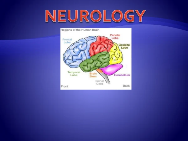
Anatomically lesions localised toWhere is the lesion? Meninges (Venus Sinus) Spinal fluid Cortex Subcortex (Basal Ganglia, Thalamus, Hypothalamus) Brain Stem (Midbrain, Pons & Medulla oblongata) Cerebellum Foramen magnum (Craniocervical Junction) Cranial Nerves Spinal Cord (ends at Lower border L1) Anterior Horn Cell Disorder Nerve root (Dorsal & Ventral) Plexus Peripheral Nerve Neuromuscular Junction Muscle
History • Instantaneous • “Electrical events” • Epilepsy • Myoclonic jerks • Neuralgic pain • Vascular events • Subarachnoid H’age (SAH) • Intracerebral H’age
History • Maximal over minutes • Vascular events • Migranous events • Maximal over Hours • Infective events • Inflammatory: GBS, Myelitis • Vascular: stroke • Vasculitic:GCA, Mononeuritis multiplex
History • Maximal over Days • Intoxication: Iatrogenic • Infection: HSV encephalitis, Meningitis • Inflammation: MS, GBS
History • Maximal over weeks/months • Brain tumours • Expanding unruptured aneurysms • Degenerative: CJD • Some polyneuropathies • Some myopathies: Steriod induced
History • Maximal over months/years • Neurodegenerative • Parkinson’s (PD) • Alzheimer’s • Cerebellar ataxias • Motor Neurone Disease (MND) • Most Neuropathies • Most myopathies
Single or multiple • Migraine • Epilepsy • TIA • Syncope • Trigeminal Neuralgia • Multiple Sclerosis (Relapsing Remitting)
Documenting Hx • No different in Neurology • Presenting complaint (PC) • Hx of the PC • Past Medical: Injuries, Psychiatric, Op, Arteriopath • Medication: Recreational use • Social/Employment: Driver, Smoker / Alcohol • Family Hx: Stroke, MND, PD, Dementias, Tremors, DM, MS
Common presenting complaints in Neurology • Funny turns • Seizures and LOC • Headaches • Dizziness & Vertigo • Confusion • Weakness of arms / legs • Abnormal movements • Loss of balance • Walking difficulties • Numbness and tingling, pins and needles • Visual failure, diplopia
HPC • As much detail as possible • When and where • Previous episodes • Witness accounts • Exacerbating and relieving factors • Treatments and changes to Rx • Associated symptoms
Recurrent attacks of LOC • Postures and manoeuvres • Drugs/Alcohol • Palpitations • Prodromal features • Post-ictal confusional states • Eye witness account • Treatments
Examination • Higher Mental Functions • Cranial Nerves • Motor • Sensory • Cerebellar • Gait • Sphincters • Skull and Spine • Neck stiffness • Neurocutaneous markers • General examination • Other systems
Higher Mental Functions • Appearance and behaviour • Mood and Affect • Thought form and content • Sensorium (GCS) and Cognition • Awareness • Sleep • Drowsiness • Stupor • Coma • Perceptual disturbances: Hallucinations • MMSE • Speech and Language
1. Olfactory 2. Optic 3. Occulomotor 4. Trochlear 5. Trigeminal 6. Abducens Smell Vision Elevate, depress and adduct, pup: constrict Depression, adduction, intorsion Face sensation, muscles of mastication Abduction Cranial nerves
7. Facial 8. Vestibulocochlear 9. Glossopharyngeal 10. Vagus 11. Spinal Accessory 12. Hypoglossal Muscles of facial expression, Anterior 2/3 tongue taste Hearing and balance Taste posterior 1/3 tongue, gag reflex Gag reflex, motor to soft palate, pharynx, larynx. Autonomic fibres to oesophagus, stomach, small intestine, heart, trachea, viscera Sternocleidomastoid, Trapezius Motor control tongue Cranial Nerves
Motor System • Bulk and nutrition • Wasting • Tone • Power • Reflexes • Babinski
DTR • 0 Absent • +/- Present with reinforcement • + Reduced • 2+ Normal • 3+ Increased Brisk Exaggerated • 4+ Pathologically brisk with clonus
Sensory System • Side to side • Proximal to distal • Pin prick • Touch • Vibration • Joint position sense • Romberg’s • Cortical sensation
Cerebellar signs • Intention Tremors • Titubation • Ataxia • Truncal ataxia • Dysdiachokinesis • Slurred speech and dysarthria • Hypotonia • Past pointing Dysmetria • Nystagmus • Tandem walking heel-toe walking • Rebound phenomenon • Pendular knee jerk • Hyporeflexia • Finger nose / Heel shin co-ordination (watch out for weakness)
Gait • Normal • Hemiplegic / Circumduction • Parkinsonian • Cerebellar • High stepping/ steppage or stamping • Waddling / Trendelenburg • Spastic • Scissor gait • Antalgic • Functional
Diagnostic tests • CSF analysis (LP) • EEG • Evoked Potentials • EMG • NCS • CT • MR • DAT • SPECT • Bloods
Typical Cerebrospinal Fluid Findings in Various Types of Meningitis Test Bacterial Viral Fungal Tubercular Opening pressure Elevated Usually normal Variable Variable WBC≥1,000 per mm3 <100 per mm3 Variable Variable Cell differential Predominance of Predominance of Predominance Predominance PMNs* lymphocytes†of lymphocytes of lymphocytes Protein Mild to marked Normal to elevated ElevatedElevated elevation CSF-to-serum glucose Normal to marked Usually normal Low Low ratio decrease CSF = cerebrospinal fluid; PMNs = polymorphonucleocytes. *—Lymphocytosis present 10 percent of the time. †—PMNs may predominate early in the course.
EEG • Encephalitis • Seizure Disorder • Encephalopathy • Anoxic brain injury • Degenerative conditions (CJD)
Trimodality EPs • Visual Evoked Responses • Brain Stem Auditory Evoked Response • Somatosensory EP
EMG/NCS • Muscle vs Motor Neuron • Demyelinative vs Axonal • Nerve root vs Plexopathy • Localisation of mononeuropathy • NMJ disorders: MG, LEMS • Entrapment Neuropathy
Neuropathy • Demyelinating • Slowed conduction • Preserved amplitude • Axonal • Reduced amplitude • Normal NCV
Neuroradiology • CT Head +/- contrast • MRI (MRA, MRV) • DWI (acute stroke) • PWI • FLAIR • MR Angiogram • PET/SPECT
Neurological Emergencies • Status Epilepticus • Coma • Traumatic Brain Injury (TBI) • Acute Stroke • Infections (Meningitis) • Subarachnoid Haemorrhage • Raised intracranial pressure Herniation • Acute Spinal cord compression • Acute Neuromuscular respiratory paralysis • Acute Visual loss • Delirium
Clinical scenario • 35 years old lady • 2/7 ago started with pins and needles in feet followed by difficulty walking then in the last 24 hours unable to hold a cup in her hands and could not get out of the bed • Past: Had diarrhoeal illness2 weeks ago. • O/E:-
O/E • Hypotonia • Faccid weakness • Areflexia • Bilateral Bell’s palsy • No UMN signs • Glove and stocking sensory disturbance • Diagnosis ??
GBS History Examination Flaccid weakness Hypotonia Hyporeflexia Cranial nerves involvement Respiratory muscle involvement Autonomic involvement Sensory disturbance No UMN signs
GBS Mortality rate 3 to 5 % Symmetric rapidly progressive, ascending, flaccid paralysis from a demyelinating poly radiculoneuropathy Post infective, post inflammatory 10% starts in ULs Progresses over the initial days up to 4 weeks Plateaux and then improves afterwards Proximal weakness Bells palsy in 50% Prior infection GIT/Resp
Diagnosis of GBS Classical history & findings Neurophysiology: Slowing of nerve conduction Serology: Campylobactor, CMV, EBV, HSV, Mycoplasma Antibodies: Anti GM1, Anti GQ1b CSF analysis: High protein with normal cells (Albumino-cytological dissociation) (? Neuro-imaging) Papilledema in GBS