Download
1 / 21
210 likes | 254 Views
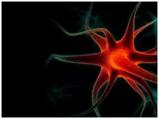
Explore cerebellar degeneration in cats through MRI images showing prominent cerebellar sulci, paucity of folia, and smaller cerebellum. Understand the impact on the brain structure.

E N D
Neurology Topic-Cerebellar Degeneration Figure 1. Sagittal and transverse T2-Weighted Images demonstrate prominent cerebellar sulci and paucity of folia diffusely involving the cerebellar hemispheres. The cerebellum appears smaller than normal. Topic-Feline Ischemic Encephalopathy Figure 1. Image 1: Dorsal T1 weighted MRI after administration of contrast. The MRI reveals a parasitic track lesion (labeled #1) extending from the right olfactory bulb to the frontal lobe. There is also enhancement along the entire parenchymal lesion into the temporal lobe, which is the pathway of the parasite through the parenchyma. The parasite exited the parenchyma over the right temporal lobe. The parasite was found in the area of the meninges on the right temporal lobe that appears enlarged after administration of contrast on the T1 weighted MR (labeled #2). Note the enhancement of the meninges over the right temporal lobe which represents the region of the infarction to the MCA. Figure 2. Ventral aspect of the brain of a cat with FIE. Note the location of the Cuterebra larva exiting the brain at the level of the right MCA and the rows of black spines on the larva. An mri of this cat’s brain revealed a parasitic track lesion extending from the right cribriform plate, through the olfactory bulb, frontal lobe and into the temporal lobe before exiting the parenchyma just rostral to the right piriform lobe and the origin of the right MCA. Topic-Head Tilt Figure 1. Left head tilt in a cat. Figure 2. MRI of same cat with left head tilt seen in previous figure. Figure 3. Paradoxical vestibular syndrome in a dog. Figure 4. Choroid plexus papilloma at postmortem.
Neurology Topic-Hydrocephalus Figure 1. Hydrocephalus: T1-weighted transverse image of the head of a dog with severe hydrocephalus. The level of the image is approximately mid-cerebrum. Fluid dilation is seen in the lateral and third ventricles. A very thin remnant of cerebral cortex can be seen lying just under the cranium (Mary O. Smith). Figure 2. Hydrocephalus: T1-weighted saggital image of the same dog as in image 1. Fluid dilation is seen in the lateral third and fourth ventricles (Mary O. Smith). Topic-Meningioma Figure 1. A 2.0 x 2.0 x 1.5cm left extra-axial parietal lobe mass. The mass is heterogeneously hyperintense on T2-WI and non-uniformly isointense on T1-WI, and homogeneously contrast enhances with evidence of a dural tail sign. The left cerebral hemisphere is swollen and there is peritumoral hyperintensity on T2-WI. There is evidence of transtentorial herniation and deviation of the midline to the right. The calvaria adjacent to the tumor is thickened and hypointense. The right frontal sinus has infiltrate that is hyperintense on T2-WI and isointense on T1-WI, and heterogeneously contrast enhances. Topic-Otitis Media and Interna Figure 1. Bilateral bulla infiltrate that is hyperintense on T2-weighted images, isointense on T1-weighted images (T1-WI) and contrast enhances. The lesion extends from the left middle ear through the petrous temporal bone to the left ventrolateral brainstem. There is a wedged shape homogenously contrast enhancing left brainstem lesion on transverse T1-WI. Topic-Schiff-Sherrington Phenomenon Figure 1. Schiff-Sherrington phenomenon in a Labrador retriever with a thoracolumbar spinal fracture. The dog was ambulatory with intact proprioception in the forelimbs, but in lateral recumbency had forelimb extension, which was resistant to manual attempts to flex the limbs (Mary Smith). Topic-Spinal Dysraphism Figure 1. The cerebellum is concave along its caudal vermal surface and the medulla is overcrowded in the caudal fossa and kinked on the sagittal T2WI. There is attenuation of the CSF pathway at the occipital foramen. There is a small accumulation of fluid in the region of the quadrigeminal cistern. There is moderate lateral ventriculomegaly. There is a moderately sized cavitary hyperintense intramedullary lesion located at the level of C1 to C6 on the sagittal T2WI.
Neurology Topic-Steroid-Responsive Meningitis- Arteritis – Dogs Figure 1. CSF cytology (cytospin preparation) from a beagle dog suffering from the acute form of steroidresponsive meningitis-arteritis. Note the neutrophilic pleocytosis. Figure 2. CSF cytology (cytospin preparation) from a beagle dog suffering from the protracted form of steroidresponsive meningitis-arteritis. Note the predominantly mononuclear pleocytosis. The numerous erythrocytes are indicative of blood contamination, which is frequently likely due to vessel damage. Figure 3. Cerebrospinal fluid from a beagle dog with steroid-responsive meningitis-arteritis. Note the turbid CSF sample. Nucleated cell count: >2,000 cells/mcl. Topic-Vestibular Disease, Geriatric – Dogs Figure 1. Typical head tilt in canine vestibular disease. Topic-Vestibular Disease, Idiopathic – Cats Figure 1. Typical head tilt in feline idiopathic vestibular disease.
Neurology Topic- Cerebellar Degeneration Figure 1. Sagittal and transverse T2-Weighted Images demonstrate prominent cerebellar sulci and paucity of folia diffusely involving the cerebellar hemispheres. The cerebellum appears smaller than normal.
Neurology Topic- Feline Ischemic Encephalopathy Figure 1. Image 1: Dorsal T1 weighted MRI after administration of contrast. The MRI reveals a parasitic track lesion (labeled #1) extending from the right olfactory bulb to the frontal lobe. There is also enhancement along the entire parenchymal lesion into the temporal lobe, which is the pathway of the parasite through the parenchyma. The parasite exited the parenchyma over the right temporal lobe. The parasite was found in the area of the meninges on the right temporal lobe that appears enlarged after administration of contrast on the T1 weighted MR (labeled #2). Note the enhancement of the meninges over the right temporal lobe which represents the region of the infarction to the MCA.
Neurology Topic- Feline Ischemic Encephalopathy Figure 2. Ventral aspect of the brain of a cat with FIE. Note the location of the Cuterebra larva exiting the brain at the level of the right MCA and the rows of black spines on the larva. An mri of this cat’s brain revealed a parasitic track lesion extending from the right cribriform plate, through the olfactory bulb, frontal lobe and into the temporal lobe before exiting the parenchyma just rostral to the right piriform lobe and the origin of the right MCA.
Neurology Topic- Head Tilt Figure 1. Left head tilt in a cat.
Neurology Topic- Head Tilt Figure 2. MRI of same cat with left head tilt seen in previous figure.
Neurology Topic-Head Tilt Figure 3. Paradoxical vestibular syndrome in a dog.
Neurology Topic-Head Tilt Figure 4. Choroid plexus papilloma at postmortem.
Neurology Topic-Hydrocephalus Figure 1. Hydrocephalus: T1-weighted transverse image of the head of a dog with severe hydrocephalus. The level of the image is approximately mid-cerebrum. Fluid dilation is seen in the lateral and third ventricles. A very thin remnant of cerebral cortex can be seen lying just under the cranium (Mary O. Smith).
Neurology Topic-Hydrocephalus Figure 2. Hydrocephalus: T1-weighted saggital image of the same dog as in image 1. Fluid dilation is seen in the lateral, third, and fourth ventricles (Mary O. Smith).
Neurology Topic-Meningioma Figure 1. A 2.0 x 2.0 x 1.5cm left extra-axial parietal lobe mass. The mass is heterogeneously hyperintense on T2-WI and non-uniformly isointense on T1-WI, and homogeneously contrast enhances with evidence of a dural tail sign. The left cerebral hemisphere is swollen and there is peritumoral hyperintensity on T2-WI. There is evidence of transtentorial herniation and deviation of the midline to the right. The calvaria adjacent to the tumor is thickened and hypointense. The right frontal sinus has infiltrate that is hyperintense on T2-WI and isointense on T1-WI, and heterogeneously contrast enhances.
Neurology Topic-Otitis Media and Interna Figure 1. Bilateral bulla infiltrate that is hyperintense on T2-weighted images, isointense on T1-weighted images (T1-WI) and contrast enhances. The lesion extends from the left middle ear through the petrous temporal bone to the left ventrolateral brainstem. There is a wedged shape homogenously contrast enhancing left brainstem lesion on transverse T1- WI.
Neurology Topic- Schiff-Sherrington Phenomenon Figure 1. Schiff-Sherrington phenomenon in a Labrador retriever with a thoracolumbar spinal fracture. The dog was ambulatory with intact proprioception in the forelimbs, but in lateral recumbency had forelimb extension, which was resistant to manual attempts to flex the limbs (Mary Smith).
Neurology Topic-Spinal Dysraphism Figure 1. The cerebellum is concave along its caudal vermal surface and the medulla is overcrowded in the caudal fossa and kinked on the sagittal T2WI. There is attenuation of the CSF pathway at the occipital foramen. There is a small accumulation of fluid in the region of the quadrigeminal cistern. There is moderate lateral ventriculomegaly. There is a moderately sized cavitary hyperintense intramedullary lesion located at the level of C1 to C6 on the sagittal T2WI.
Neurology Topic- Steroid-Responsive Meningitis-Arteritis - Dogs Figure 1. CSF cytology (cytospin preparation) from a beagle dog suffering from the acute form of steroid-responsive meningitis-arteritis. Note the neutrophilic pleocytosis.
Neurology Topic- Steroid-Responsive Meningitis-Arteritis - Dogs Figure 2. CSF cytology (cytospin preparation) from a beagle dog suffering from the protracted form of steroid-responsive meningitis-arteritis. Note the predominantly mononuclear pleocytosis. The numerous erythrocytes are indicative of blood contamination, which is frequently likely due to vessel damage.
Neurology Topic-Steroid-Responsive Meningitis-Arteritis - Dogs Figure 3. Cerebrospinal fluid from a beagle dog with steroid-responsive meningitis-arteritis. Note the turbid CSF sample. Nucleated cell count: >2,000 cells/mcl.
Neurology Topic-Vestibular Disease, Geriatric - Dogs Figure 1. Typical head tilt in canine vestibular disease.
Neurology Topic-Vestibular Disease, Idiopathic - Cats Figure 1. Typical head tilt in feline idiopathic vestibular disease.