Download

1 / 10
230 likes | 1.06k Views
Preparing a Blood Smear. Samples for Hematology. Capillary blood and venous blood can be used for hematology tests. Capillary blood is beneficial if a small amount of blood is needed. Venous blood via venipuncture is beneficial if a larger amount of blood is needed for testing.

E N D
Samples for Hematology • Capillary blood and venous blood can be used for hematology tests. • Capillary blood is beneficial if a small amount of blood is needed. • Venous blood via venipuncture is beneficial if a larger amount of blood is needed for testing. • Most hematology tests are run using blood collected with the anticoagulant, ethylenediaminetetraacetic acid, or EDTA for short. • EDTA is the anticoagulant found in lavender top tubes.
Samples for Hematology: CBC • A complete blood count or CBC is one of the most commonly ordered test in the hematology department. • When running a CBC there are several separate tests run. • Tests included in a CBC are…. • Red Blood Cell Count (RBCs) • White Blood Cell Count (WBCs) • Hemoglobin (Hgb) • Hematocrit (Hct) • Mean Corpuscular Volume (MCV) • Mean Corpuscular Hemoglobin (MCH) • Mean Corpuscular Hemoglobin Concentration (MCHC) • Differential count (Diff) • Platelet Count (PLTs) • Blood Cell Morphology
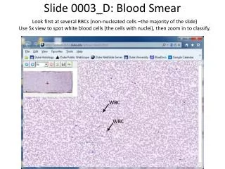
Blood Smears • A CBC also includes an examination of a blood smear if there are issues with the CBC. • A smear of blood is made and then stained and examined by the laboratory personnel. • Stained slides can be read manually (by a person) and sometimes by a machine. • Ultimately a person must review the slides before results are reported. • Blood smears are checked for types of WBCs, shapes of RBCs, and the presence of platelets. • Morphology the structure and shape of cells • The morphology off all cells are reviewed.
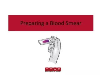
Preparing a Blood Smear • Equipment needed to perform a blood smear include….. • Clean slides (prewashed or cleaned) • Capillary supplies or Venipuncture supplies • Fresh tube of blood
Preparing a Blood Smear • Making a blood smear • Place a small drop of blood ½ inch from the frosted end of a slide. • The end of a second slide is used to spread the blood drop out on the first slide. • Pull the end of the slide through the blood drop toward you and then push in the opposite direction forcing the blood in your direction.
Preparing a Blood Smear • Making a blood smear • A good smear should….. • cover around ¾ of the slide • have a feathered edge • move from thick to thin • cells should be evenly distributed • RBCs should not overlap • Factors that affect the smear include….. • blood drop size • angle of the slide used to spread • speed of the procedure • Making an acceptable slide takes lots of practice!
Preparing a Blood Smear • Staining the blood smear • The smear should be stained as soon as possible after the slide dries. • If the slide cannot be stained immediately, it can be immersed in methanol (fixed) and stained later. • Blood smears are most commonly stained with Wright’s stain. • Wright’s stain can come in many forms including one dip stains, two stains, multiple stains, or automated staining of slides.
Reading a Blood Smear • Reading a Blood Smear • The elements you will be looking for in a stained blood smear are RBCs, WBCs, and PLTs. • The WBCs will be counted according to their type. • RBCs and PLTs are easier to identify than WBCs. • To categorize the different types of WBCs, look for the following things….. • Size in comparison to the RBCs • Nuclear shape and staining • Cytoplasm makeup and staining
Reading a Blood Smear • Reading a Blood Smear • You count each WBC in each category until you reach a total of 100 cells. • The number you got for each type of cell divided by 100 will give you the percentage each type of cell is found in each sample. • For example if you counted….. • 55 neutrophils = 55% of WBCs were neutrophils • 30 lymphocytes = 30% of WBCs were lymphocytes • 10 monocytes = 10% of WBCs were monocytes • 3 eosinophils = 3% of WBCS were eosinophils • 2 basophils = 2% of WBCS were basophils