The Trigeminal Nerve
The Trigeminal Nerve. Dr. Nimir Dr. Safaa. Objectives List the trigeminal nerve nuclei and their location Follow up the course of trigeminal nerve from its point of central connections to exit and down to its target areas.


The Trigeminal Nerve
E N D
Presentation Transcript
The Trigeminal Nerve Dr. Nimir Dr. Safaa
Objectives • List the trigeminal nerve nuclei and their location • Follow up the course of trigeminal nerve from its point of central connections to exit and down to its target areas. • Describe the sensory and motor components of the trigeminal nerve.
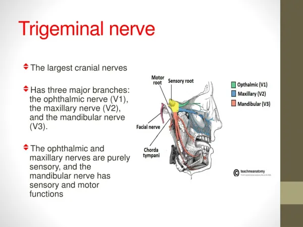
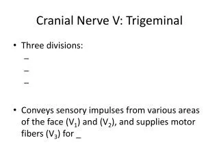
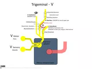
Trigeminal Nerve (Cranial Nerve V): • It is the largest cranial nerve and contains both sensory and motor fibers. • It is sensory to greater part of head and motor to several muscles including muscles of mastication.
Trigeminal Nerve Nuclei: • It has four nuclei: • (1) Main sensory nucleus. • (2) Spinal nucleus. • (3)Mesencephalic nucleus. • (4) Motor nucleus.
Main sensory nucleus lies in posterior part of pons lateral to the motor nucleus. • Spinal nucleus continuous superiorly with main sensory nucleus and extends inferiorly through medulla oblongata and into upper part of spinal cord as far as second cervical segment.
Mesencephalic Nucleus composed of unipolar cells situated in lateral part of gray matter around cerebral aqueduct. • It extends inferiorly into pons as far as main sensory nucleus. • Motor nucleus is situated in ponsmedial to main sensory nucleus.
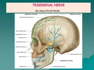
Course of the Trigeminal Nerve • Trigeminal nerve leaves anterior aspect of pons as a small motor root and a large sensory root. • It passes forward out of posterior cranial fossaand rests on apex of petrousbone in middle cranial fossahere sensory root expands to form trigeminal ganglion. • Ophthalmic, maxillary, and mandibularnerves arise from the anterior border of ganglion.
Ophthalmic nerve (V1) contains only sensory fibers leaves skull through superior orbital fissure to orbital cavity. • Maxillary nerve (V2) also contains onlysensoryfibers leaves the skull through foramen rotundum. • Mandibular nerve (V3) contains both sensory and motor fibers and leaves skull through foramen ovale. • The sensory fibers to skin of face from each division supply a distinct zone with little or no overlap of dermatomes .
Sensory Components of the Trigeminal Nerve • Pain, temperature, touch, and pressure from skin of face and mucous membranes travel along axons whose cell bodies are situated in the trigeminal ganglion. • The central processes of these cells form sensory root of trigeminal nerve. • About half the fibers divide into ascending and descending branches when they enter the pons. • The remainder ascend or descend without division.
The ascending branches terminate in main sensory nucleus, and descending branches terminate in spinal nucleus. • Touch and pressure are conveyed by nerve fibers that terminate in the main sensory nucleus. • Pain and temperature pass to spinal nucleus.
Proprioceptive impulses from muscles of mastication and from facial and extraocular muscles are carried by fibers of unipolar cells of the mesencephalic nucleus that have bypassed trigeminal ganglion. • Axons of the neurons in the main sensory and spinal nuclei and central processes of cells in mesencephalic nucleus now cross median plane and ascend as trigeminal lemniscusto terminate on nerve cells of ventral posteromedialnucleus of the thalamus. • Axons of these cells now travel through genu internal capsule to postcentralgyrus (areas 3, 1, and 2) of the cerebral cortex.
Motor Component of the Trigeminal Nerve • Motor nucleus receives corticonuclear fibers from both cerebral hemispheres. • It also receives fibers from reticular formation, red nucleus, tectum, and medial longitudinal fasciculus. • In addition, it receives fibers from the mesencephalic nucleus, thereby forming a monosynaptic reflex arc. • Cells of motor nucleus give rise to axons that form motor root. • Motor nucleus supplies muscles of mastication, tensor tympani, tensor velipalatini, mylohyoidand anterior belly of digastricmuscle.