Overview: Microbial Model Systems Viruses called bacteriophages
0.5 m. Figure 18.1. Overview: Microbial Model Systems Viruses called bacteriophages Can infect and set in motion a genetic takeover of bacteria, such as Escherichia coli. E. coli and its viruses
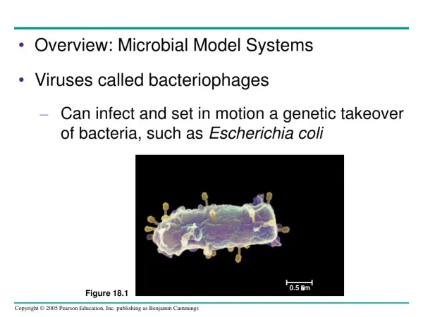

Overview: Microbial Model Systems Viruses called bacteriophages
E N D
Presentation Transcript
0.5 m Figure 18.1 • Overview: Microbial Model Systems • Viruses called bacteriophages • Can infect and set in motion a genetic takeover of bacteria, such as Escherichia coli
E. coli and its viruses • Are called model systems because of their frequent use by researchers in studies that reveal broad biological principles • Beyond their value as model systems • Viruses and bacteria have unique genetic mechanisms that are interesting in their own right
Virus Bacterium Animalcell 0.25 m Animal cell nucleus Figure 18.2 • Recall that bacteria are prokaryotes • With cells much smaller and more simply organized than those of eukaryotes • Viruses • Are smaller and simpler still
Concept 18.1: A virus has a genome but can reproduce only within a host cell • Scientists were able to detect viruses indirectly • Long before they were actually able to see them
Figure 18.3 The Discovery of Viruses: Scientific Inquiry • Tobacco mosaic disease • Stunts the growth of tobacco plants and gives their leaves a mosaic coloration
In the late 1800s • Researchers hypothesized that a particle smaller than bacteria caused tobacco mosaic disease • In 1935, Wendell Stanley • Confirmed this hypothesis when he crystallized the infectious particle, now known as tobacco mosaic virus (TMV)
Structure of Viruses • Viruses • Are very small infectious particles consisting of nucleic acid enclosed in a protein coat and, in some cases, a membranous envelope
Viral Genomes • Viral genomes may consist of • Double- or single-stranded DNA • Double- or single-stranded RNA
Capsomereof capsid RNA DNA Capsomere Glycoprotein 18 250 mm 70–90 nm (diameter) 20 nm 50 nm (b) Adenoviruses Figure 18.4a, b (a) Tobacco mosaic virus Capsids and Envelopes • A capsid • Is the protein shell that encloses the viral genome • Can have various structures
Membranousenvelope Capsid RNA Glycoprotein 80–200 nm (diameter) 50 nm (c) Influenza viruses Figure 18.4c • Some viruses have envelopes • Which are membranous coverings derived from the membrane of the host cell
Head DNA Tail sheath Tail fiber 80 225 nm 50 nm (d) Bacteriophage T4 Figure 18.4d • Bacteriophages, also called phages • Have the most complex capsids found among viruses
General Features of Viral Reproductive Cycles • Viruses are obligate intracellular parasites • They can reproduce only within a host cell • Each virus has a host range • A limited number of host cells that it can infect
VIRUS DNA Entry into cell and uncoating of DNA Capsid Transcription Replication HOST CELL Viral DNA mRNA Viral DNA Capsid proteins Self-assembly of new virus particles and their exit from cell Figure 18.5 • Viruses use enzymes, ribosomes, and small molecules of host cells • To synthesize progeny viruses
Reproductive Cycles of Phages • Phages • Are the best understood of all viruses • Go through two alternative reproductive mechanisms: the lytic cycle and the lysogenic cycle
The Lytic Cycle • The lytic cycle • Is a phage reproductive cycle that culminates in the death of the host • Produces new phages and digests the host’s cell wall, releasing the progeny viruses
1 Attachment. The T4 phage usesits tail fibers to bind to specificreceptor sites on the outer surface of an E. coli cell. 2 Entry of phage DNA and degradation of host DNA.The sheath of the tail contracts,injecting the phage DNA intothe cell and leaving an emptycapsid outside. The cell’sDNA is hydrolyzed. 5 Release. The phage directs productionof an enzyme that damages the bacterialcell wall, allowing fluid to enter. The cellswells and finally bursts, releasing 100 to 200 phage particles. Phage assembly 3 Synthesis of viral genomes and proteins. The phage DNAdirects production of phageproteins and copies of the phagegenome by host enzymes, usingcomponents within the cell. 4 Assembly. Three separate sets of proteinsself-assemble to form phage heads, tails,and tail fibers. The phage genome ispackaged inside the capsid as the head forms. Head Tail fibers Figure 18.6 Tails • The lytic cycle of phage T4, a virulent phage
The Lysogenic Cycle • The lysogenic cycle • Replicates the phage genome without destroying the host • Temperate phages • Are capable of using both the lytic and lysogenic cycles of reproduction
Phage DNA The phage attaches to a host cell and injects its DNA. Many cell divisions produce a large population of bacteria infected with the prophage. Phage DNA circularizes Phage Occasionally, a prophage exits the bacterial chromosome, initiating a lytic cycle. Bacterial chromosome Lytic cycle Lysogenic cycle Certain factors determine whether The bacterium reproduces normally, copying the prophage and transmitting it to daughter cells. The cell lyses, releasing phages. Prophage Lytic cycle is induced Lysogenic cycle is entered or New phage DNA and proteins are synthesized and assembled into phages. Phage DNA integrates into the bacterial chromosome,becoming a prophage. Figure 18.7 • The lytic and lysogenic cycles of phage , a temperate phage
Reproductive Cycles of Animal Viruses • The nature of the genome • Is the basis for the common classification of animal viruses
Table 18.1 • Classes of animal viruses
Viral Envelopes • Many animal viruses • Have a membranous envelope • Viral glycoproteins on the envelope • Bind to specific receptor molecules on the surface of a host cell
Glycoproteins on the viral envelope bind to specific receptor molecules(not shown) on the host cell, promoting viral entry into the cell. Capsid RNA 2 1 Envelope (with glycoproteins) Capsid and viral genome enter cell 3 HOST CELL The viral genome (red) functions as a template forsynthesis of complementary RNA strands (pink) by a viral enzyme. Viral genome (RNA) Template 5 mRNA Complementary RNA strands also function as mRNA, which is translated into both capsid proteins (in the cytosol)and glycoproteins for the viral envelope (in the ER). New copies of viral genome RNA are made using complementary RNA strands as templates. 4 Capsid proteins ER Copy of genome (RNA) Glyco- proteins 6 Vesicles transport envelope glycoproteins to the plasma membrane. 7 New virus 8 A capsid assembles around each viral genome molecule. Figure 18.8 • The reproductive cycle of an enveloped RNA virus
RNA as Viral Genetic Material • The broadest variety of RNA genomes • Is found among the viruses that infect animals
Glycoprotein Viral envelope Capsid RNA(two identicalstrands) Reversetranscriptase Figure 18.9 • Retroviruses, such as HIV, use the enzyme reverse transcriptase • To copy their RNA genome into DNA, which can then be integrated into the host genome as a provirus
1 Reverse transcriptase catalyzes the synthesis of a DNA strand complementary to the viral RNA. 2 The virus fuses with the cell’s plasma membrane. The capsid proteins are removed, releasing the viral proteins and RNA. Membrane of white blood cell HIV Reverse transcriptase catalyzes the synthesis ofa second DNA strand complementary to the first. 3 Reverse transcriptase HOST CELL Viral RNA 4 The double-stranded DNA is incorporated as a provirus into the cell’s DNA. RNA-DNAhybrid 0.25 µm HIV entering a cell DNA ChromosomalDNA NUCLEUS Provirus 5 Proviral genes are transcribed into RNA molecules, which serve as genomes for the next viral generation and as mRNAs for translation into viral proteins. RNA genomefor the nextviral generation mRNA 6 The viral proteins include capsid proteins and reverse transcriptase (made in the cytosol) and envelope glycoproteins (made in the ER). 7 Capsids are assembled around viral genomes and reverse transcriptase molecules. Vesicles transport the glycoproteins from the ER to the cell’s plasma membrane. 8 9 New viruses bud off from the host cell. Figure 18.10 New HIV leaving a cell • The reproductive cycle of HIV, a retrovirus
Evolution of Viruses • Viruses do not really fit our definition of living organisms • Since viruses can reproduce only within cells • They probably evolved after the first cells appeared, perhaps packaged as fragments of cellular nucleic acid
Concept 18.2: Viruses, viroids, and prions are formidable pathogens in animals and plants • Diseases caused by viral infections • Affect humans, agricultural crops, and livestock worldwide
Viral Diseases in Animals • Viruses may damage or kill cells • By causing the release of hydrolytic enzymes from lysosomes • Some viruses cause infected cells • To produce toxins that lead to disease symptoms
Vaccines • Are harmless derivatives of pathogenic microbes that stimulate the immune system to mount defenses against the actual pathogen • Can prevent certain viral illnesses
Emerging Viruses • Emerging viruses • Are those that appear suddenly or suddenly come to the attention of medical scientists
(b) The SARS-causing agent is a coronavirus like this one (colorized TEM), so named for the “corona” of glycoprotein spikes protruding from the envelope. (a) Young ballet students in Hong Kong wear face masks to protect themselves from the virus causing SARS. Figure 18.11 A, B • Severe acute respiratory syndrome (SARS) • Recently appeared in China
Outbreaks of “new” viral diseases in humans • Are usually caused by existing viruses that expand their host territory
Figure 18.12 Viral Diseases in Plants • More than 2,000 types of viral diseases of plants are known • Common symptoms of viral infection include • Spots on leaves and fruits, stunted growth, and damaged flowers or roots
Plant viruses spread disease in two major modes • Horizontal transmission, entering through damaged cell walls • Vertical transmission, inheriting the virus from a parent
Viroids and Prions: The Simplest Infectious Agents • Viroids • Are circular RNA molecules that infect plants and disrupt their growth
Originalprion Prion Many prions Normalprotein Newprion Figure 18.13 • Prions • Are slow-acting, virtually indestructible infectious proteins that cause brain diseases in mammals • Propagate by converting normal proteins into the prion version
Concept 18.3: Rapid reproduction, mutation, and genetic recombination contribute to the genetic diversity of bacteria • Bacteria allow researchers • To investigate molecular genetics in the simplest true organisms
The Bacterial Genome and Its Replication • The bacterial chromosome • Is usually a circular DNA molecule with few associated proteins • In addition to the chromosome • Many bacteria have plasmids, smaller circular DNA molecules that can replicate independently of the bacterial chromosome
Replicationfork Origin of replication Termination of replication Figure 18.14 • Bacterial cells divide by binary fission • Which is preceded by replication of the bacterial chromosome
Mutation and Genetic Recombination as Sources of Genetic Variation • Since bacteria can reproduce rapidly • New mutations can quickly increase a population’s genetic diversity
EXPERIMENT Researchers had two mutant strains, one that could make arginine but not tryptophan (arg+ trp–) and one that could make tryptophan but not arginine (arg trp+). Each mutant strain and a mixture of both strains were grown in a liquid medium containing all the required amino acids. Samples from each liquid culture were spread on plates containing a solution of glucose and inorganic salts (minimal medium), solidified with agar. Mixture Mutantstrainarg+trp– RESULTS Only the samples from the mixed culture, contained cells that gave rise to colonies on minimal medium, which lacks amino acids. Figure 18.15 • Further genetic diversity • Can arise by recombination of the DNA from two different bacterial cells Mutantstrainargtrp+
Mixture Mutantstrainarg+trp– Mutantstrainarg–trp+ No colonies(control) No colonies(control) Coloniesgrew CONCLUSION Because only cells that can make both arginine and tryptophan (arg+ trp+ cells) can grow into colonies on minimal medium, the lack of colonies on the two control plates showed that no further mutations had occurred restoring this ability to cells of the mutant strains. Thus, each cell from the mixture that formed a colony on the minimal medium must have acquired one or more genes from a cell of the other strain by genetic recombination.
Mechanisms of Gene Transfer and Genetic Recombination in Bacteria • Three processes bring bacterial DNA from different individuals together • Transformation • Transduction • Conjugation
Transformation • Transformation • Is the alteration of a bacterial cell’s genotype and phenotype by the uptake of naked, foreign DNA from the surrounding environment
Phage DNA B+ A+ 1 Phage infects bacterial cell that has alleles A+ and B+ Host DNA (brown) is fragmented, and phage DNA and proteins are made. This is the donor cell. A+ 2 B+ Donorcell 3 A bacterial DNA fragment (in this case a fragment withthe A+ allele) may be packaged in a phage capsid. A+ Crossingover Phage with the A+ allele from the donor cell infects a recipient A–B– cell, and crossing over (recombination) between donor DNA (brown) and recipient DNA (green) occurs at two places (dotted lines). 4 A+ A– B– Recipientcell The genotype of the resulting recombinant cell (A+B–) differs from the genotypes of both the donor (A+B+) and the recipient (A–B–). 5 A+ B– Figure 18.16 Recombinant cell Transduction • In the process known as transduction • Phages carry bacterial genes from one host cell to another
1 m Sex pilus Figure 18.17 Conjugation and Plasmids • Conjugation • Is the direct transfer of genetic material between bacterial cells that are temporarily joined
The F Plasmid and Conjugation • Cells containing the F plasmid, designated F+ cells • Function as DNA donors during conjugation • Transfer plasmid DNA to an F recipient cell
3 1 2 4 F Plasmid Bacterial chromosome F+ cell F+ cell Mating bridge Bacterial chromosome F+ cell F– cell A cell carrying an F plasmid(an F+ cell) can form amating bridge with an F– celland transfer its F plasmid. A single strand of the F plasmid breaks at a specific point (tip of blue arrowhead) and begins tomove into the recipient cell. As transfer continues, the donor plasmid rotates(red arrow). DNA replication occurs inboth donor and recipientcells, using the single parental strands of the F plasmid as templates to synthesize complementary strands. The plasmid in the recipient cell circularizes. Transfer and replication result in a compete F plasmid in each cell. Thus, both cells are now F+. (a) Conjugation and transfer of an F plasmid from an F+ donor to an F– recipient Figure 18.18a • Conjugation and transfer of an F plasmid from an F+ donor to an F recipient
Chromosomal genes can be transferred during conjugation • When the donor cell’s F factor is integrated into the chromosome • A cell with the F factor built into its chromosome • Is called an Hfr cell • The F factor of an Hfr cell • Brings some chromosomal DNA along with it when it is transferred to an F– cell
2 1 Hfr cell F+ cell F factor The circular F plasmid in an F+ cellcan be integrated into the circularchromosome by a single crossoverevent (dotted line). The resulting cell is called an Hfr cell (for High frequency of recombination). B+ D+ C+ C+ Hfr cell A+ D+ A+ B+ D+ A+ C+ B+ A+ A+ D+ C+ B+ B+ C– C– C– F– cell B– D– D– A+ B– D– B– A– A– A– C– B+ D– B– A– 3 4 6 Since an Hfr cell has all the F-factor genes, it can form a mating bridge with an F– cell and transfer DNA. 5 The mating bridgeusually breaks well before the entire chromosome andthe rest of the F factor are transferred. A+ The location and orientation of the F factor in the donor chromosome determine the sequence of gene transfer during conjugation. In this example, the transfer sequence for four genes is A-B-C-D. A single strand of the F factorbreaks and begins to move through the bridge. DNA replication occurs in both donor and recipient cells, resulting in double-stranded DNA Temporary partial diploid Recombinant F– bacterium C– C– B+ B– D– D– B+ B– A– A– A+ A+ 7 Two crossovers can result in the exchange of similar (homologous) genes between the transferred chromosome fragment (brown) and the recipient cell’s chromosome (green). 8 The piece of DNA ending up outside thebacterial chromosome will eventually be degraded by the cell’s enzymes. The recipient cell now contains a new combination of genes but no F factor; it is a recombinant F– cell. (b) Conjugation and transfer of part of the bacterial chromosome from an Hfr donor to an F– recipient, resulting in recombination • Conjugation and transfer of part of the bacterial chromosome from an Hfr donor to an F– recipient, resulting in recombination Figure 18.18b