Download
1 / 48
510 likes | 974 Views
Chapter 22. Anatomy & Physiology Fifth Edition Seeley/Stephens/Tate (c) The McGraw-Hill Companies, Inc. The Lymphatic System and Immunity. The human body recognizes anything other than its own as an invader.

E N D
Chapter 22 Anatomy & Physiology Fifth Edition Seeley/Stephens/Tate (c) The McGraw-Hill Companies, Inc.
The Lymphatic System and Immunity • The human body recognizes anything other than its own as an invader. • When these foreign bodies are capable of living in the human body and are harmful, they are called pathogens. • Plasma contains antibodies against cells other than its own. These antibodies are formed when the infants are about three months old. Recall when the blood types were formed. • Thus, there is the lymphatic system in the body to identify and destroy foreign bodies. (by forming antibodies) • But, how do they distinguish our own from the others? • When the reaction is against each specific foreign body, it is called immune response or immunity.
Organization and Functions of the Lymphatic System • In the lymphatic system there are: • Lymphatic fluid, lymph, that contains lymphocytes • Vessels that transport lymph • And sites where large contents of lymphocytes are held: lymph nodes, spleen, and thymus. • Functions of the lymphatic system • The lymph system has three major functions: • 1. Fluid Balance. Circulating blood release about 30 L of fluid into interstitial space each day. Of the 27 L are returned to the circulation. The remaining 3L will enter the lymphatic system as lymph. The lymph passes through the lymphatic system and enter back to the blood vessels. In addition to water, lymph contains substances as in plasma and substances extracted from cells. • 2. Fat Absorption. Fats and other substances are absorbed from the digestive tract and carried through the lymphatic system. • 3. Defense. Immune system (lymphocytes, B-cells, T-cells)
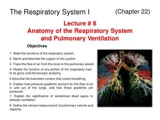
Lymphatic Vessels • Lymphatic vessels are spread out throughout the body except in the CNS (Fig 22.1) • They originate from within lymphocyte producing organs and empty into the thoracic duct. • They contain many lymphatic nodes and nodules. • Like blood vessels, they have small vessels called lymphatic capillaries which lay close to the blood capillaries. • Lymph soluble fluid will be picked up through the capillaries and pushed back to larger truck. • They even have valves to control the flow of lymph through very low pressure of fluid.
Lymphatic Organs • Diffuse lymphatic tissue • Dispersed lymphocytes, macrophages and other cells. No clear boundaries. (Fig. 22.2) • Lymphoid Nodules • Packed with lymphocytes. • Flexible in size depending upon the number of lymphocytes • Spread in loose C.T. of the digestive, respiratory and urinary systems. Larger patches in intestinal system call Peyer’s patches. • In lymph nodes they are called follicles, there they contain a germinal center, where lymphocytes divide. • Though filled with lymphocytes, lymphoid nodules could be infected: tonsillitis, appendicitis.
Tonsils • Large groups of lymph nodules and diffuse lymphatic tissue located in the oral cavity (Fig. 22.3) • Protection against bacteria, etc.. In the nose and mouth. • Three types of tonsils: pharyngeal, palatine, and lingual • Enlarged pharyngeal tonsil is called adenoid. • Lymph Nodes ( Fig. 22.1 and Fig. 22.4) • Found throughout the body except in the brain. • They are encapsulation of lymphocytes, macrophages and reticular cells with blood vessels • 1-25mm in diameter and are subdivided into two regions called, a medulla and cortex. • Within the cortex germinal centers are found. This is where lymphocyte division takes place. • Act as sieve and removal of 99% of antigens while reactivating T- cells and B-cells.
The Spleen • Upper and posterior part of the abdominal cavity. • Largest of the lymphatic tissue, 160g. (Fig. 22.5) • Contains two types of lymphatic tissue: white pulp (arterial supply) and red pulp (venous supply). • The spleen detects and responds to foreign substances in the blood, destroys worn-out RBC’s and acts as a blood reservoir.
Thymus Gland • On top of the heart • T-cells mature here • Becomes large during the first or second year. • Intrinsic size is largest at puberty and then decreases. • Produces lymphocytes which then move to other lymphatic tissues. • Blood-thymic barrier - reticular cells wrap around capillaries and prevent large molecules from entering the cortex of the thymus.
Immunity • How the human body defends itself against damage from foreign substances such as microorganisms and harmful chemical as toxins. • Innate (non-specific) and adaptive (specific) immunity. These two types are distinguished by the way they respond to specific stimulations and how they memorize the events. • Specificity. Innate immunity can act against bacteria in genera. (no memory) • Memory. Adaptive immunity can distinguish among different kinds of bacteria and generally get more sensitive with each new encounter.
Innate Immunity • Mechanical Mechanism • Physical barriers, such as the skin and membranes • Chemical Mediators (Table 22.1) • Molecules which contribute to develop immunity. • Kill bacteria: lysozyme and sebum mucus • Others: histamine and kinins by vasodilation; interferon production, etc.. • Complement • Is a group of approximately 20 proteins that makes up approximately 10% of the globulin part of serum. They are a group of proteins activated in the form of a cascade and provide protection by attacking the bacterial membrane. They attach to and form holes in the membranes. The proteins can also attach to bacteria membrane and stimulate macrophages to phagocytize the bacteria.
Interferons: proteins that protect the body against viral infection and possibly cancer. • Cells for Innate Immunity • Review the major functions of, neutrophils, monocytes, macrophages, basophils, mast cells, eosinophils, and natural killer cells. (Table 22.2) • Inflammatory Response • Tissue damage caused by bacteria or others may induce inflammation of tissue as it releases histamine, prostaglandins, kinins, etc… • Vasodilation attracts chemotatic phagocytes and other leukocytes to the region as well as fibrinogen to form fibrin in order to localize damage.
Adaptive Immunity • Substances to stimulate adaptive immunity are antigens, of which molecule weights could be as large as 10,000 or more. (produce antibodies) • Haptens are small molecules capable of combining with larger molecules to stimulate adaptive immunity response. • Two type of antigens: foreign antigens (allergen) are from outside of the body, and self antigens (auto-immune) are molecules of its own body. • For example. Allergic reaction is by foreign antigens, while autoimmune disease is from self antigens.
Immunity has been divided into two types: humoral (body fluid) immunity and cell mediated immunity. • Specificity : recognition of antigen • The specificity is established because of the specific receptors located on the surface of T and B cells. • Versatility: there are many antigens. • And there are many different forms of lymphocytes that are made against them. • Memory: the adaptive immune system has a memory. With the presence of the foreign body, lymphocytes responding to it begin to initiate cell divisions. • The presence of antigen leads to the formation of active and memory cells. • The active cells respond to antigens, while the memory cells wait until the next onslaught. • In this manner, the second response to the same antigen will be fast.
Lymphocytes • 25% of circulating white cells. • The majority of lymphocytes are in peripheral tissues. • Types of lymphocytes • We have already seen that there are three types of lymphocytes. (Fig. 22.9) • T cells: 80% of circulating lymphocytes (cell mediated immunity) • B cells: 10-15% , plasma cells produce antibodies (immunoglobulins) and react antigenic pathogens. ( antibody-mediated immunity) • Natural killer cell: the remainder: they attack foreign cells, normal cells infected with viruses and cancer cells. They immunologically survey peripheral tissues.
Cytotoxic T cells attack foreign cells or cells infected with viruses. Provide cell-mediated immunity. • Helper T cells stimulate the activities of T cells and B cells. • Suppressor T cells inhibit T cells and B cells.
Origin and development of lymphocytes • Lymphocytes travel around the entire body and have significantly long life span. • 80% survive up to 4 years and some to 20 years. • As we have seen: lymphocytes are made in the red bone marrow and some continue to develop in the thymus. (Fig. 22.9) • Pre-B cells and pre-T cells are in the red bone marrow. Pre-B cells mature in the red bone marrow into B cells. T cells mature in the thymus.
A positive selection process keep cells capable of immune response. Those which are incapable will die. • Each group of B or T cells capable of responding to a specific antigen is a clone • Each clone is capable of responding to a particular antigen and there are many different clones. • When the clones respond to self-antigens, negative selection eliminates such clones. • Most of this process occurs during prenatal development, but continues throughout life.
Activation of Lymphocyte • Lymphocytes are made in response to a specific antigen and in a large quantity. • Antigenic Determinants and Antigens Receptors • An antigen may have many antigenic determinants (epitopes) (Fig. 22.10) to which lymphocytes can respond. • Each antigenic determinate can activate a specific lymphocyte. Thus is possible that an antigen with many epitopes can activate many different (clones) lymphocytes. • Each lymphocyte from the same clone will have the same antigen receptor. • For example, a t cell receptor has a variable and constant regions. (Fig. 22.11). The variable region will have specific antigen binding sites. Thus a clone of T cells can bind a specific antigenic determinant. The other T cells within the same clone could have different antigen binding sites. • The B-cell receptor is similar, but larger.
Major Histocompatibility Complex Antigens • Some are direct, but most lymphocyte activation involves glycoproteins on the surface of cells called MHC molecules. All cell membranes have MHC> • MHC class I molecules(Fig. 22.12a) • On nucleated cells, foreign or self proteins are fragmented in the nucleated cells and become antigens. • They combine with MHC class I molecule in the cell and the complex are transported on the surface of the cell. • At the surface of the cell membrane, the foreign antigen/MHC I complex will attract T cells and the cell will be destroyed. (cell mediated immunity), while self antigen/MHC I will not.
MHC class II Molecules(Fig 22.12b) • The more complex and advanced lymphocytes stimulation use MHC II molecules. • These molecules are found in antigen-presenting cells, such as B cells, macrophages, monocytes and dendritic cells. • Antigen-presenting cells phagocytically ingest unprocessed antigens, process them and let them combine with MCH II molecules. • the complex will be presented on the surface of the cells. • They stimulate the other (lymphocytes) immune cells to divide and to cause the destruction of the antigen.
Costimulation • In addition to the interaction between the presented MHC II/ antigen and antigen receptor, costimulation, with cytokinins for example, is needed (Fig.22 13a) • Another costimulation (Fig. 23. 13b) is achieved cross linking two cells with other molecules such as CD4, B7, CD28, etc. • Other example of cytokinins are listed in Table 22.4. Note interferons and interleukins. • Helper T Cells • Enhances more production of T and B cells • On activation helper T cells produce a variety of cytokins that coordinated specific and nonspecific defenses and stimulate cell mediated immunity. • Study Fig. 22.14 and 22.15 to find how proliferation of helper T cell can be achieved. • In short, with the help of cytokinins, such as interleukins, the number of helper T cells increase and thus stimulate B cells or effector T cells, which release perforin, produces hole in infected cells.
Inhibition of lymphocytes • Inhibition of lymphocytes proliferation against its own self-antigen is achieved by tolerance. • Deletion of self-reactive lymphocytes • During prenatal development when immature lymphocytes are exposed to the self antigens, they die. Thus, no lymphocytes which respond to the self antigen will be found as the subject matures. • Preventing activation of lymphocytes • By lack of costimulation • Activation of suppresser T cell. • This is not well understood. The suppressor T cells release suppressive cytokins.
Antibody-mediated Immunity • When exposed to an antigen, the body activates B cells and produces antibodies. • the antibodies are found in body fluids thus respond to extracellular antigens. • Antibodies • Antibodies are produced in B cells in response to an antigen and are found in plasma. • Plasma proteins have four major components: albumin, alpha, beta and gamma globulins. • Antibodies are found in gamma globulins group, thus sometimes are called gamma globulins or immunoglobulins (Ig). • There are five types (Table 22.5) • IgG, IgM, IgA, IgE, and IgD
They all consist of four peptides. (Fig 22.16) connected with disulfide bonds and have a constant region and a antigen binding variable region. • The constant region may attach to cells, such as macrophages, basophils, etc.. • Effects of Antibodies • The function of antibody is to find antigen and destroy it. • Neutralization, agglutination and precipitation, activation of complement. Attraction of phagocytes, enhancement of phagocytes, and stimulation of inflammation. • Antibodies can counteract the action of antigens in several ways (Fig. 22.17) • Upon binding with an antigen, the antibody inactivates the antigen. • Inactivation may lead to co-precipitation of antigen and antibodies. • Binding of two may stimulate phagocytic activities and the release of inflammation chemicals.
Antibody production • The primary Response • The first encounter with an antigen, an B cell divides and differentiate leading to antibodies production (Fig. 22.18a). • Antibodies, IgM and IgD, are on the surface of B cells. • B cells, which are small lymphocytes, activated by antigen starts cell divisions. • Some become large plasma cells, which produce antibodies and other become small memory B cells. • The primary process takes 1-14 days, the antigen may cause tissue damage during this period.
The secondary or memory response • When the immune system is exposed to the same antigen after the primary response • The memory cells quickly divide to produce plasma cells and large quantity of antibody is released, providing immune protection. • the response is quick, hours to a few days, and the the quantity of antibody production is large. • Good defense against the disease. • The memory cells, which may survive for years, are also formed.
Cell-mediated Immunity • It is done by T cells and is against the intracellular attack, microorganism, virus, because antigens in cells are presented. • Activation of T cells to antigens is regulated by antigen-presenting cells and helper T cells like. B cells, activated T cells produce cytotoxic T cells and memory T cells (Fig 22.19) • Cytotoxic T cells • They lyse cells and produce cytokines that produce inflammation and phagocytosis.\ • Viral antigens, tumors antigens and foreign antigens on the surface of cell are the stimulants to cytotoxic T cells. • Released cytokins may recruit macrophages for phagocytosis and inflammation.
Immune Disorders • Autoimmune disorders • Identify own body cells as antigens, such as rheumatoid arthritis. • Some viruses, measles and influenza, have a sequence similar to those of myelin proteins. Thus, antibodies which attack viruses may also attack myelin sheath. • Unusual types of MHV proteins. • Allergies • Excessive immune response to antigens. • Immune interactions