Chapter 15 Hypersensitivity Reaction
Chapter 15 Hypersensitivity Reaction . I . Hypersensitivity reaction II. Type I Anaphylactic Reactions III. Type II Antibody-mediated cytotoxic hypersensitivity IV. Type III Immune complex-mediated hypersensitivity

Chapter 15 Hypersensitivity Reaction
E N D
Presentation Transcript
Chapter 15 Hypersensitivity Reaction I .Hypersensitivity reaction II. Type I Anaphylactic Reactions III. Type II Antibody-mediated cytotoxic hypersensitivity IV. Type III Immune complex-mediated hypersensitivity V. Type IV T cell-mediated hypersensitivity
I .Hypersensitivity reaction 1. Concept 2. Types of Hypersensitivity Reaction
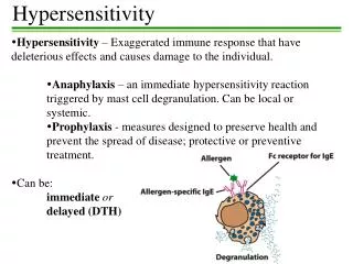
1. Concept Under some circumstances, immunity, rather than providing protection, produces damaging and sometimes fatal results. Such deleterious reactions are known collectively as hypersensitivity reactions, but it should be remembered that they differ from protective immune reactions only in that they are exaggerated or inappropriate and damaging to the host. The cellular and molecular mechanisms of the two types of reaction are virtually identical.
2. Types of Hypersensitivity Reaction Hypersensitivity reaction were divided into four classes, designated types I-, by Gell and Coombs Type I: Anaphylactic Reactions Type II: Antibody-mediated cytotoxic hypersensitivity Type III: Immune complex-mediated hypersensitivity Type IV: T cell-mediated hypersensitivity
II. Type I: Anaphylactic Reactions 1. Concept of Type I 2. Characteristics of type I 3. Mechanism of type I a. Sensitization phase b. Activation phase c. Effect phase 4. Clinical aspects of type I
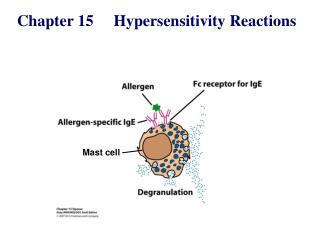
1. Concept of Type I Anaphylactic reaction are mediated by IgE antibodies, which bind to receptors on mast cells and basophils. When cross-linked by antigen, the IgE antibodies trigger the mast cells (basophils) to release several pharmacologically active agents that are responsible for the characteristic symptoms of anaphylaxis. Reactions are rapid, occurring within minuts after challenge, i.e. exposure to antigen.
2. Characteristics of type I a. Anaphylactic reaction are rapid, occurring within minutes after challenge, exposure to same antigen. It reaches its peak within 10-15 minutes; then it fades without leaving any residual damage, if it isn’t fatal. b. IgE antibodies-mediated and IgG4(a little), without IgA, IgM, IgG1,2,3 and complements. c.The mast cells and basophils are the main effectors of type I reaction, and the many pharmacologically active agents released by the mast cells and basophils. d. The symptoms of anaphylaxis seen in systemic reactions. This would induce a typical wheal and flare reaction consisting of blood vessel dilation and increase in permeability and induce difficulty in breathing because of constriction of bronchiolar muscles, uterine cramps, or involuntary urination and defecation. e. The different individual have distinct difference.
3. Mechanism of type I a. Sensitization phase b. Activation phase c. Effect phase - preformed mediators - Newly synthesized mediators
Allergen Allergen TCR MHC-II CD4 Th cell IL-4 mast cell mast cell B m B cell Plasma cell FcR Sensitized IgE Degranulation Mechanism: Smooth muscle cell, Small blood vessel, Mucous gland, Blood platelets, Sensory nerve endings, Eosinophil, And so on.
Principal mediators involved in type I hypersensitivity Mediator Activities primary 1. Histamine: Increased vascular permeability; Smooth muscle contraction. 2. Serotonin: Increased vascular permeability; Smooth muscle contraction. 3. Eosinophil chemotactic factor: (ECF-A) Eosinophil chemotaxis 4. Neutrophil chemotactic factor: (NCF-A) Neutrophil chemotaxis 5. Proteases: Degradation of blood-vessel basement membrane; Generation of complement split products.
Principal mediators involved in type I hypersensitivity Mediator Activities Secondary 1. Platelet-activating Platelet aggregation and degranulation. Factor:(PAF) Contraction of pulmonary smooth muscles. 2. Leukotrienes: Increased vascular permeability; ( SRS-A)Contraction of pulmonary smooth muscles. 3. Prostaglandins: Vasdilation; Contraction of pulmonary smooth muscles. Platelet aggregation. 4. Brady Kinin: Increased vascular permeability; Smooth-muscles contraction.
4. Clinical aspects of type I a. Anaphylactic shock ( drug anaphylactic reaction) b. Anaphylactic reaction of respiratory system ( bronchial asthma ) c. Allergic dermatitis d. Intestinal allergy
III. Type II: Antibody-mediated cytotoxic hypersensitivity 1. Concept of type II 2. Characteristics of type II 3. Mechanism of type II 4. Clinical aspects of type II
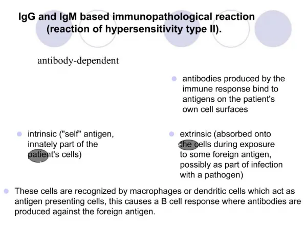
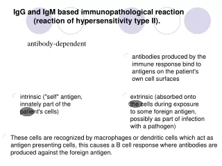
1. Concept of type II A typeII hypersensitivity reaction (cytotoxic reaction) occur when IgM or IgG antibodie bind to antigen on the surface of cells and activate the complement cascade, leading to cell damage or de ath through complement-mediated lysis or Ab-dependent cell-mediated cytotoxicity(ADCC).
2. Characteristics of type II a. IgM and IgG antibodies-mediated b. In type II hypersensitivity binding of the appropriate antibody directly to an antigen on the surface of a cell produces damage to that cell through a variety of mechanisms. c. The mechanisms of type II hypersensitivity involve either the complete complement sequence and eventual lysis of the cell or opsonic effects mediated by receptors for Fc or C3b, which lead to phagocytosis and destruction of the cell by macrophages, neutrophils and NKC (ADCC).
Cell-surface Ag Fc 1. IgG Target cell Target cell NK 3. Cytotoxic action (ADCC) CR 2. Phagocyte Antibody C Complement mediated lysis 3. Mechanism of type II
4. Clinical aspects of type II a. Transfusion reactions b. Rh incompatibility reaction* c. Drug-induced reaction d. Autoimmune type II hypersensitivity -- Autoimmune hemolytic anemia -- Goodpasture’s syndrome -- Anti-receptor auto-antibody disease -- Anti-hormone auto-antibody disease
First birth postportum subsequent pregnancy RhD mother Bcell Anti-Rh IgM RhD red cells Anti-RhD lysis RhD fetus RhD fetus Haemolytic disease of the newborn
IV. Type III Immune complex-mediated hypersensitivity 1. Concept of type III 2. Characteristics of type III 3. Mechanism of type III 4. Clinical aspects of type III
1. Concept of type III A type III hypersensitivity --Immune complex reaction--occur when complexes of antigen and IgM or IgG antibody accumulate in the circulation or in tissue andactivate the complement cascade. Granulocytes are attracted to the site of activation, and damage results from the release of lytic enzymes from their granules. Reaction occur within hours of challenge with antigen.
2. Characteristics of type III a. IgM and IgG-mediated b. When antigen and antibody meet at the appropriate concentrations, they form insoluble antigen-antibody complexes(IC). c. IC can activate the complement cascade. Release of certain products of complements (C3a and C5a) causes a local increase in vessel permeability and permits the release of serum (edema) and chemotactic attraction of neutrophils. The neutrophils release degradative lysosomal enzymes that produce the tissue damage characteristic of these reaction. d. IC (the circulating complexes) accumulate in or near vessel walls and deposit in such tissues as kidney , joints, or skins. If the site of reaction is a vessel well, the outcome is hemorrhage and necrosis; If the site is a glomerular basement membrane, loss of integrity and release of protein and red blood cells into the urine results; and if the site is a joint meniscus, destruction of synovial membrane and cartilage occurs.
3. Mechanism of type III a. Immune complexes production and deposition Immune complexes size and deposition The amounts of antibodies and deposition The sites of deposition of the complexes b. The mechanisms of damage
Antigen+antibody Immune complexes Platelets Complement Macrophages Anaphylatoxins lysis Microthrombi Vasoactive amines Activation and release of IL-1 and reactive Oxygen intermediates Mast cell Attract Neutrophils Vasoactive amines Release granular contents Increased vascular permeability Vasodilation 3. Mechanism of type III
4. Clinical aspects of type III a. Arthus’ reaction b. Serum sickness c. Infection-associated syndrome - glomerulonephritis - Rheumatic fever - Rheumatoid arthritis - Other infectious diseases
V. Type IV: T cell-mediated hypersensitivity 1. Concept of type IV 2. Characteristics of type IV 3. Mechanism of type IV 4. Clinical aspects of type IV
1. Concept of type IV Cell-mediated immunity reaction(CMI)--also called delayed-type hypersensitivity(DTH)--is mediated by T cells rather than by antibody. Upon activation, the T cells release cytokines that cause accumulation and activation of macrophages, which, in turn release lysosomal enzymes that cause local damage. This type of reaction has a delayed onset and may occur 1-2days after challeage with antigen.
2. Characteristics of type IV a. CMI, when activated by contact with an antigen presented by APC, T cells release soluble mediators, cytokines, some of which attract and activate other mononuclear cells such as monocytes, macrophages, and nonimmune lymphocytes. b. This type of reaction has a delayed onset and may occur 1-2 days after challenge with Ag. c. The reaction reveals that the mononuclear infiltrates appear as a perivascular cuff before extensively invading the site of deposition of antigen. Later reaction show a more comlex pattern, with the arrival of B cells and the formation of granuloma in persistent lesions. d. The reaction have not apparent individual difference.
antigen T cell Inflammatory mediators cytokines Activated macrophages 3. Mechanism of type IV
4. Clinical aspects of type IV a. Contact Sensitivity ( skin inflammatory reaction) b. Allograft Rejection c. Graft Versus Host Reaction (GVHR) d. Cell-mediated Hypersensitivity Reaction