The Case
Evaluation of the Sudden and Severe Headache: Diagnosis and Management Michael Gerardi, MD, FAAP, FACEP Vice-Chairman, Department of Emergency Medicine Morristown Memorial Hospital Director, Pediatric Emergency Medicine Children’s Medical Center Morristown, New Jersey. The Case.

The Case
E N D
Presentation Transcript
Evaluation of the Sudden and Severe Headache:Diagnosis and ManagementMichael Gerardi, MD, FAAP, FACEPVice-Chairman, Department of Emergency MedicineMorristown Memorial HospitalDirector, Pediatric Emergency MedicineChildren’s Medical CenterMorristown, New Jersey
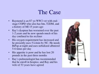
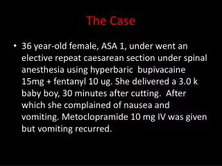
The Case One hour prior to ED presentation, a 42 year old man was jogging and “hit” by the worst headache of his life. It was associated with some nausea and the feeling as if he was going to pass out. He rested for 30 minutes but the headache persisted as a diffuse, throbbing pain radiating to the base of his skull.
The Case (Continued) EMS was called. The patient felt as if he could not concentrate, there was no confusion, nor was there any other focal neurologic complaint. There was no past medical history, no medications, no family history, and no significant use of alcohol, tobacco or other drugs.
If a patient presented with the worst headache of his life, what is the work-up that should be initiated? a. Non-contrast CT b. LP after neg. CT c. LP without CT d. CT, LP, and angiography
Objectives • What is the differential of a “thunderclap headache”? • What is the sensitivity of neuroimaging in subarachnoid hemorrhage (SAH)? • What constitutes a “positive” lumbar puncture in SAH and when should it be performed? • Do patients with suspected SAH who have a negative CT and lumbar puncture require additional imaging to “rule-out” expanded but unruptured aneurysm?
Headache • 1 of 10 top presenting complaints • 1 to 2% of visits to ED • 18 million outpatient visits • 638 million days of work lost per year • 78% of women and 64% of men had experienced at least one in the prior year • 36% of women and 19% men suffer from recurrent headaches
Headache • Most have primary headache disorders • migraine • tension • Only a few have treatable secondary causes that threaten life, limb, brain such as subarachnoid hemorrhage • 1 - 4 % of headache visits
“Worst” Headache • Normal exam: 12- 33% SAH • Abnormal exam: 25% SAH • Initial hemorrhage may be fatal • Early definitive surgery improves outcomes • Patients with greatest likelihood of benefiting from surgery are most likely to receive incorrect diagnosis
Physicians Consistently Misdiagnose SAH 1. Failure to appreciate spectrum of clinical presentation 2. Failure to understand limitations of CT 3. Failure to perform and correctly interpret the results of LP
ED Goals in Headache Patients 1. Differentiate life-threatening from benign 2. Initiate prompt treatment 3. Provide prompt pain relief 4. Prevent drug seeking and refer 5. Minimize resource utilization in ED 6. Optimize patient use of ED 7. Increase pre-ED treatment and reduce ED use
Differential Diagnosis of Headache • Onset • Location • Associated symptoms • Pain characteristics • Duration • Prior history • Diagnostic tests • Physical exam
Medical Conditions That Present With Headache • Pheochromocytoma • Hyperthyroidism • SLE • Giant Cell Arteritis • Fibromyalgia
Types of Headaches in the ED Final Diagnosis Percentage Infection - not intracranial 39.3 Tension HA 19.3 Miscellaneous 14.9 Post-traumatic 9.3 Hypertension related 4.8 Vascular (Migraine) 4.5 No diagnosis 6.0 SAH 0.9 Meningitis 0.6
Ped HA Compared to Literature: Serious Conditions Author # Age Tumor Bleed Meningitis Burton 288 2-18 0 0 0.3 Fodden 106 0-90 4.7 8.5 0 Leicht 485 15-89 2.7 1.0 0.8 Dopeshi 872 2-92 0.1 0.9 0.6 Dickman 124 16-65 0 0 0
Causes of Headache That Require Specific Therapy • Subarachnoid hemorrhage • Meningitis • Encephalitis • Cervicocranial-artery dissection • Temporal arteritis • Acute angle-closure glaucoma • Hypertensive emergency
Causes of Headache That Require Specific Therapy • Carbon Monoxide poisoning • Pseudotumor cerebri • Cerebral venous and dural sinus thrombosis • Acute stroke (hemorrhagic or ischemic) • Mass Lesion • tumor • abscess • intracranial hematoma • parameningeal infection
Headache Danger Signals • Onset • after 40 years • new or different headache • subacute HA that worsens • exertion, sex, coughing, straining • Worst ever experienced
Headache Danger Signals: Associated With Neurologic Change • Memory impairment • Ataxia • Drowsiness • Sensory loss • Signs of meningeal irritation
Headache Danger Signals: Associated With Neurologic Change • Progressive visual or neurologic change • Confusion • Weakness • Loss of coordination • Asymmetry of pupils, DTRs
Headache Danger Signals: Abnormal Medical Evaluation • Fever • Chronic malaise • Arthralgia • HTN • Myalgia • Wt loss • Tender, poorly pulsatile temporal arteries
Subarachnoid Hemorrhage • 10% of all acute CVAs • 30,000 persons/year • 10 -16/100,000 • 1% of all ED patients with acute cephalgia
Subarachnoid Hemorrhage • Incidence of 16 /100,000 • about 33,600 cases per year • 54% secondary to ruptured aneurysm • Without treatment, 40% of aneurysm pts. have recurrent bleeding • Aneurysm pt who survives initial rupture and is treated conservatively: • 50% survival at one year
Subarachnoid Hemorrhage • Onset: Acute • Location: Global • Ass Sx: N,V, meningismus, focal • Pain: Worst ever • Duration: Brief • Prior Hx: No • Dx tests: CT 80-90% • Phys ex: Focal signs, LOC, meningismus
Subarachnoid Hemorrhage • Warning leaks in 50% • CT misses up to 10% small leaks • Suspect if: • > 35 years • no previous HA • no fading of HA • came on with exertion • altered LOC or neuro deficits • stiff neck
Subarachnoid Hemorrhage: Neurologic Findings • Sudden HA without localizing findings • Altered mentation • Confusion, lethargy • Bilateral extensor plantar reflex • Unusual to find focal deficits
Causes of Non-Traumatic Subarachnoid Hemorrhage • “Berry” aneurysms • AVM • Cerebral angiomas • Mycotic aneurysm • Extension from parenchymatous hemorrhage • Anticoagulation therapy
Causes of Non-Traumatic Subarachnoid Hemorrhage • Systemic bleeding diathesis • Hemorrhagic encephalitis • Hemorrhagic cerebral vasculitis • Hemorrhage into CNS tumors or metastases • Unknown
Intracranial Aneurysms • Women: men = 3 : 2 • 4 million Americans • 20% multiple aneurysms • Increase in mid-20s • Peak incidence of 12% by age 60 • Risk of spontaneous rupture 1 to 3%/yr • Peak 40 to 60 years
Arteriovenous Malformations • 10-15% of SAH • Spontaneous hemorrhage • Any age but usually < 30 • Incidence 3% per year • Incidence of major neurologic deficit or mortality: 50%
Conditions Associated with Cerebral Aneurysm Development • HTN • Polycystic kidney disease • Connective tissue disorders • Coarctation of aorta • Pregnancy induced HTN • Family history of CVAs • Bacterial endocarditis
Warning Headache • 20 - 50% patients with SAH have HA days or weeks before index episode • unusually severe • distinct • “Thunderclap” headache • Day and Raskin 1996 • intense, acute, peak intensity at onset • develop in seconds • maximal intensity in minutes • lasts hours to days
“Thunderclap” Headache • 25% associated with SAH • “Warning” headache • followed by SAH in 5% to 60% • Expansion or dissection of unruptured aneurysm • Cerebral venous thrombosis • Exertional / coital headache
Subarachnoid Hemorrhage: Morbidity and Mortality 28,000 ruptured aneurysms 10,000 18,000 dead/disabled available for Rx 3,000 7,000 8,000 10,000 died rapidly misdiagnosed dead or functional no warning or missed disabled survivors
Misdiagnosis of Symptomatic Cerebral Aneurysm: Mayer 1996 • 217 patients with symptomatic SAH • 54 / 217 misdiagnosed • 46 / 217 minimal findings • viral meningitis 15% • migraine 13% • uncertain etiology 13% • Failure to consider SAH
Missed Cerebral AneurysmsMayer 1996 • 9 / 43 (21%) CTs initially read as neg. • 6 of these 9 : (+) SAH • 48% re-bleed or deteriorated (vs. 2%) • Good or excellent outcomes • 91% initially correct • 53% if misdiagnosed
SAH…But not “Classic” • Roughly half have minor bleeding with atypical features • Nonstrenuous activities (34%) • Sleep (12%) • HA in any location (localized, generalized, mild) • May be relieved by non-narcotic analgesics • Diagnosed as migraine, tension-type, sinusitis
SAH…But not “Classic” • Prominent neck pain • Cervical sprain, arthritis • Confusion, agitation, restless • psychiatric diagnoses • Syncope / trauma • Traumatic SAH • Syncope / abnormal ECG • “MI and then trauma” • 91% SAH have cardiac dysrhythmias and ECGs mimicking ischemia
SAH: Most patients have... • Abrupt onset of severe, unique headache, or neck pain • Abnormal findings on neurologic examination • Subtle meningismus or ocular findings
International Headache Society • A first episode of severe headache cannot be classified as migraine: • more than 4 episodes • nor as tension-type headache: • more than 9 episodes • First or worst headache requires evaluation • as do qualitatively different headaches
Can a CT Scan Safely “Rule Out” SAH? • First diagnostic study • Thin cuts ( 3 mm) through base of brain • Blood on CT function of Hgb • Hgb < 10: blood isodense • Sensitivity decreases over time from onset of symptoms
Acute HA of Recent OnsetLeido A. Headache 1994 • 27 patients; 24 - 77 yo • 1 hr to 13 days after HA onset • no previous similar HA • no focal neurologic signs • all had CT; LP if CT neg
Acute HA of Recent OnsetLeido A. Headache 1994 • 9 of 27 (33%) : SAH • 4 (+) CT • 5 normal CT, (+) LP • 2 of 19 LPs: meningitis • CT scanning should be done with first severe acute headache
CT & Subarachnoid Hemorrhage:Sames et al: 1996 Sensitivity of NGCT: Group 1 (symptoms < 24 hrs) 93.1% Group 2 (symptoms > 24 hrs) 83.8% “A normal NGCT does not reliably exclude the need for LP”
SAH: CT SensitivitySames: Acad Emerg Med Jan 1996 • 181 patients; aged 13-86 with SAH • Sensitivity 91.2% • pain < 24 hrs 93.1% • pain > 24 hrs 83.8% • LP 100% sensitive if neg CT • “A normal NGCT does not reliably exclude the need for LP”
SAH Diagnosis: LP NeededSidman: Acad Emerg Med Sep 1996 • 140 patients; aged 10-88 • Sensitivity of CT • < 12 hrs 80/80 100% • > 12 hrs 49/60 81.7% • Overall, 11/140 had (-) CT and (+) LP • overall sensitivity 92.1%
Morgenstern LB, et al:Worst headache and SAH: Prospective, modern CT and spinal fluid analysis.Ann Emerg Med Sept 1998. • 38,730 patients over 16 months, prospectively screened for “worst HA” • Blinded neuroradiologists • Neg CT LP • cell count x 2 • visual and spectrophotometric detection of xanthochromia • CSF D-dimer assay
Morgenstern, et al: Ann Emerg Med 1998 • 455 headaches & 107 “worst headache” • CT: 18 of 107 (17%): (+) SAH • (-) CT/ (+) SAH: Only 2 (2.5%) • (95% CI, 0.3%to 8.8%) • Modern CT is sufficient to exclude 98% of SAH in patients
Morgenstern, et al: Ann Emerg Med 1998 (107 “Worst HA’s) Variables CT-/LP- CT+ CT-/LP+ Photophobia 45 28 50 Stiff neck 26 37 100 Nausea 65 36 100 Lethargy 17 40 50 Time < 24 h 58 75 50 Migraine 20 11 0 Headache 48 27 0
CT is Normal: Do LP? Yes!
What about LP First? • Duffy et al; 1982: 55 patients who underwent LP as initial w/u • Condition deteriorated immediately in 7 patients • Hillman et al; 1986: 4 alert patients with SAH who deteriorated after lumbar puncture • Both studies: • clots on CT or a dilated pupil