Download
1 / 57
580 likes | 1.07k Views
Chapter 18. Prenatal Development and Diseases Associated with Pregnancy. Learning Objectives (1 of 2). Explain process of fertilization; implantation; early development of ovum; origin of decidua, fetal membranes, and placenta

E N D
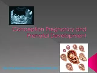
Chapter 18 Prenatal Development and Diseases Associated with Pregnancy
Learning Objectives (1 of 2) • Explain process of fertilization; implantation; early development of ovum; origin of decidua, fetal membranes, and placenta • Describe formation and elimination of amnionic fluid; conditions leading to abnormal levels • Explain causes and effects of spontaneous abortion and ectopic pregnancy • Identify and explain problems following failure of contraceptive pills or intrauterine device • Describe mechanism and clinical manifestations associated with abnormal attachment of placenta
Learning Objectives (2 of 2) • Differentiate: Identical vs. fraternal twins • Describe determination of zygosity from examination of placenta; disadvantages of a twin pregnancy • Classify types of gestational trophoblast disease, methods of treatment • Explain pathogenesis, clinical manifestations, diagnostic criteria, treatment for hemolytic disease of the newborn
Fertilization • Union of sperm and ovum occurs in fallopian tube • Sperm travel via own propulsion and by passive transported upward into the fallopian tubes by rhythmic contractions of uterine muscles • Ovum is expelled from follicle at ovulation • Fertilization is possible when sperm are present in fallopian tubes at the time egg is expelled at ovulation • First cell division completed 30 hours after fertilization • Only 1 sperm can enter egg • Sperm penetration causes zona pellucida to become impermeable to penetration by other sperm
Early Development: Fertilized Ovum • Fertilization occurs in fallopian tube • Sperm contain genetic material and enzymes for penetration • Zygote develops into a small ball of cells • Fluid accumulates to form blastocyst • Inner cell mass: forms embryo • Trophoblast: forms placenta and membranes • Blastocyst begins to differentiate • Implantation by end of 1st week • Blastocyst implants in endometrium • Amnionic sac and yolk sac form • Small germ disk and yolk sac project into chorionic cavity • Organ systems begin to form; embryo becomes cylindrical by 4th week
In Vitro Fertilization and Embryo Transfer • Some women ovulate normally but are infertile because fallopian tubes obstructed by scarring or removed due to previous ectopic pregnancies • Patient’s follicle is aspirated by laparoscopy • Ovum is fertilized and allowed to develop outside the body into 8- or 16-cell stage • Fertilized ovum implanted into uterus • Low success rate • Possibility of chromosomally abnormal embryos • Abnormal embryos usually unable to survive and are aborted
Stages of Prenatal Development • Pre-embryonic period: First 3 weeks after fertilization • Blastocyst becomes implanted and inner mass cell differentiates into 3 germ layers to eventually form specific tissues within embryo • Embryonic period: 3rd through 7th week • Begins to assume a human shape • All organ systems are formed • Very critical period of development • Fetal period: 8th week to term • Fetus continues to grow • No major changes in basic structure • Subcutaneous fat accumulates; fills body shortly before delivery
Progressive changes in fetal size,3.5 months, 4.5 months, and 5.5 months
Duration of Pregnancy • Gestation: total duration of pregnancy from fertilization to delivery • Dated from time of conception: 38 weeks • Dated from first day of last menstrual period (date of ovulation unknown): 40 weeks • First day of the calculation is two weeks before the date of conception • May be expressed as 280 days • Also expressed as 10 lunar (28-day) months or 9 calendar (31-day) months; divided into 3 periods called trimesters
Decidua, Fetal Membranes, Placenta • Decidua: endometrium of pregnancy • Decidua basalis: under chorionic vesicle • Decidua capsularis: over chorionic vesicle • Decidua parietalis: lines rest of the uterus • Chorion laeve: superficial smooth chorion • Chorion frondosum: bushy chorion • Amnionic sac: enclosed within chorion, forms a protective environment • Yolk sac: forms intestinal tract Chorionic vesicle
Placenta • Double circulation of blood • Fetoplacental circulation: from fetus to villi • Uteroplacental circulation: maternal blood circulates around villi • No actual intermixing of maternal and fetal blood • Fetus connected to placenta by umbilical cord • Functions of the placenta • Provides O2 and nutrition for fetus • Has endocrine function: synthesizes hormones (estrogen; progesterone; protein hormones) • Human placental lactogen, HPL • Human chorionic gonadotropin, HCG
Amnionic Fluid (1 of 2) • Produced by filtration and excretion • Filtration from maternal blood early in pregnancy • Fetal urine later in pregnancy • Fetus swallows fluid: • Absorbed from fetal intestinal tract into fetal circulation • Transferred across placenta into mother’s circulation • Excreted by mother in her urine • Balance is maintained in secretion and excretion of amnionic fluid • Quantity varies with stage of pregnancy
Amnionic Fluid (2 of 2) • Polyhydramnios: increased volume of amniotic fluid • Fetus unable to swallow and fluid accumulates (anencephaly) • Fluid is swallowed but not absorbed due to congenital obstruction of fetal upper intestinal tract • Oligohydramnios: reduced volume of amniotic fluid • Fetal kidneys failed to develop and no urine is formed • Congenital obstruction of urethra does not allow urine to form amnionic fluid
Gestational Diabetes • Hyperglycemia: harmful to fetus • Pregnancy hormones induce maternal insulin resistance • Diabetes results from inability to increase insulin secretion to compensate for increased insulin resistance • Diagnosis: similar to non-gestational diabetes • Diabetes usually relents following delivery
Spontaneous Abortion • 10-20% of all pregnancies • Early abortion results from • Chromosome abnormalities • Defective implantation • Maldevelopment of fetus • Late abortion results from • Detachment of placenta • Obstruction of blood supply through cord • Complication: Disseminated intravascular coagulation • Cocaine abuse: disturbs blood flow to placenta and may cause placental abruption and intrauterine fetal death
Small well-formed spontaneously aborted fetus near the end of the first trimester
Fetus spontaneously aborted late in pregnancy because of interruption of blood supply through the umbilical cord
Ectopic Pregnancy • Development of embryo outside the uterine cavity • Most common site: fallopian tubes • Predisposing factors • Previous infection of fallopian tubes • Failure of normal muscular contractions of tubal wall • Both fallopian tunes predisposed • Consequences • Rupture of fallopian tube • Profuse bleeding from torn vessels • Potentially life-threatening to mother
Ectopic Pregnancy, fallopian tube with mass of placental tissue; embryo within intact amnionic sac
Artificial Contraception • Failure of oral contraceptives • Developing embryo exposed to synthetic estrogen and progestin compounds • Exposure to estrogen and progestin compounds may induce congenital abnormalities in developing embryo • Failure of an intrauterine device • IUD predisposes pregnant uterus to infection • Must be removed as soon as pregnancy is diagnosed
Abnormal Attachment of Umbilical Cord • Velamentous insertion • Cord attached to fetal membranes than placenta • May tear or is compressed during labor • May be fatal to infant • No adverse effect on mother Velamentous insertion of umbilical cord with vessels traversing fetal membranes
Abnormal Attachment of Placenta • Normally, placenta attaches high on the anterior or posterior uterine wall • Placenta previa: placenta attached at lower part of uterus; may cover cervix • Central placenta previa: placenta covers entire cervix • Partial placenta previa: margin of placenta covers cervix • Causes episodes of bleeding late in pregnancy • Hazardous to both mother and infant • Requires delivery by cesarean section
Twins (1 of 3) • Disadvantages of twinning • Twins smaller than single infant at comparable stage of gestation • Over-distention of uterus promotes premature onset of labor • Delivery of premature infants • Reduced chance of survival • Congenital malformations occur twice as often in twins
Twins (2 of 3) • Twin transfusion syndrome • Vascular anastomoses connect placental circulations of identical twins • One twin is polycythemic and one is anemic • Tolerated if minor disproportions in blood, if severe, may be fatal to both twins • Vanishing twin: one of the twin dies and is resorbed • Blighted twin: one of the twin dies and persists as degenerated fetus
Twins (3 of 3) • Fraternal twins: 2 separate ova fertilized by 2 different sperm • Identical twins: single fertilized ovum splits • Conjoined twins: variable union between identical twins
Stages at which formation of identical twins can occur, and types of placenta associated with each stage of twinning.
A comparison of the formation of the placentas and fetal membranes in twins
Placenta of identical twins with amnions removed revealing interconnecting blood vessels
Conjoined twins exhibiting a large congenital defect in the abdominal wall
X-ray of conjoined twins demonstrating extreme curvature of fetal spine
Gestational Trophoblast Disease (1 of 4) • Trophoblastic cells covering villi continue to grow at excessive rate, producing higher HCG than in normal pregnancy • Proliferating trophoblastic tissue may invade uterus, vagina, distant sites • Hydatidiform mole (“hydatid”: fluid-filled vesicle; “mole”: shapeless structure) • Occurs in 80% of affected patients • Complete mole • Results from abnormal fertilization of an ovum lacking chromosomes and ovum fertilized by a single sperm bearing an X chromosome that is duplicated to form 46
Gestational Trophoblast Disease (2 of 4) • Hydatidiform mole: complete mole • Both X chromosomes come from the father • No embryo develops • Chorionic villi become cystic structures resembling mass of grapes (complete mole) • Hydatidiform mole: partial mole • Normal ovum fertilized by 2 sperm, resulting in a fertilized ovum with 3 sets of chromosomes (69 chromosomes) • Embryo forms but does not survive • Less likely to exhibit aggressive behavior
Gestational Trophoblast Disease (3 of 4) • Invasive mole • Trophoblastic tissue invades deeply into uterine wall • Occurs in 15% of affected patients • Aggressive, destructive
Gestational Trophoblast Disease (4 of 4) • Choriocarcinoma • May arise following incomplete removal of invasive or incompletely removed mole • Masses of proliferating trophoblast may extend into vagina • Metastasizes to lungs and brain • Treatment: curettage, periodic determination of HCG; hysterectomy; chemotherapy
Erythroblastosis Fetalis (1 of 2) • Pathogenesis • 1. Sensitization of mother to a blood group antigen in fetal RBCs • 2. Mother forms antibodies that cross placenta • 3. Maternal antibodies damage fetal RBCs • 4. Fetus increases blood production to compensate for increased RBC destruction • Variable severity • Hydrops fetalis • Less intense hemolytic process • Mild disease
Erythroblastosis Fetalis (2 of 2) • Hydrops fetalis • Severe anemia causes heart failure and impaired hepatic plasma protein synthesis • Results in edema • Hemolytic process is extremely severe, causing death • Infant dies in uterus during last trimester • Less intense hemolytic process • Infant is born alive but moderately or severely anemic • Mild disease • Infant appears normal at birth then becomes anemic and jaundiced, develops edema
Fetal Hydrops, stillborn with marked edema.Swollen abdomen from enlarged liver and spleen and fluid accumulation in peritoneal cavity.
Rh Hemolytic Disease (1 of 3) • Most cases: Rh-negative mother and Rh-positive infant • Consists of a series of allelic genes that determine multiple Rh antigens on red cells • Rh-positive: red cells contain D (Rho) antigen • May be homozygous (genotype DD) • May be heterozygous (genotype Dd) • Rh-negative: red cells lack D (Rho)antigen • Genotype dd
Rh Hemolytic Disease (2 of 3) • Mother sensitized to “foreign” antigen in infant’s cells and forms anti-D antibodies that cross placenta into infant’s blood • Rarely occurs in first pregnancy • First Rh-positive infant born to Rh-negative mother is normal • Rh antibodies not yet formed • Treatment • Exchange transfusion • Fluorescent light therapy for hyperbilirubinemia • Intrauterine fetal transfusion
Rh Hemolytic Disease (3 of 3) • Prevention • Rh immune globulin administered to mother • Contains gamma globulin with Rh antibody • Given within 72 hours after delivery of Rh-positive infant • Rh antibody coats Rh antigen sites on surface of fetal red cells in maternal circulation to reduce sensitization • Not 100% effective: 1.5% still form antibodies • Some physicians recommend injections late in pregnancy and after delivery to reduce incidence of sensitization