Download
1 / 22
220 likes | 307 Views
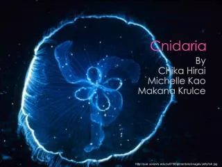
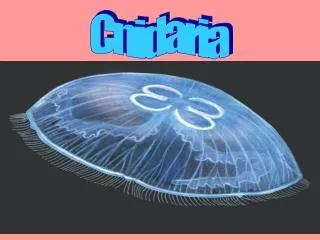
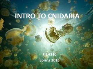
INTRO TO CNIDARIA. FISH310 Spring 2013. Defining Characteristics. secretion of complex intracellular organelles called cnidea All have basic radial symmetry Planula larvae - free-swimming, flattened, ciliated, bilaterally symmetric larva

E N D
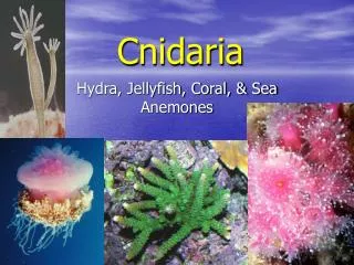
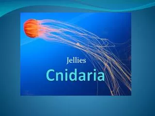
INTRO TO CNIDARIA FISH310 Spring 2013
Defining Characteristics • secretion of complex intracellular organelles called cnidea • All have basic radial symmetry • Planula larvae - free-swimming, flattened, ciliated, bilaterally symmetric larva • Possess only 2 layers of living tissue (epidermis and gastrodermis) with mesoglea in between • All have tentacles around the mouth • A single opening to the digestive system.
Hydrozoans • ~3,000 mostly marine species • Typically small with polyp and medusa stage • Members of this class include: Hydroida, Siphonophora, Hydrocorallina • Gastrodermis lacks cnidea • No cells in mesoglea
Portuguese Man O’War! http://video.nationalgeographic.com/video/manowar_portuguese
Hydrozoans • Examine colonial polyp • Try to identify species using dichotomous key • Note colony form, polymorphism, life history
Anthozoans Anthozoan Evolution! • Never have a medusa stage • Today we will look at the external and internal anatomy of a sea anemone
Sea Anemone’s at War! Acrorhagi • Hollow spherical protrusions covered with potent nematocyts • Function in defending territory Today we will see these defensive structures in our Anthopleura dissections
Cnidea (nettle/stinging thread) • Multiple Functions: food collection, defense and locomotion. • Can be specialized for wrapping around small objects, sticking to surfaces, penetrating surfaces, or secreting proteinaceous toxins. • 3 major categories of cnidea: • Spirocysts – anthozoans (corals/sea anemones) • Ptychocysts – tube anemones • Nematocysts – most common and well studied (over 30 types; many types in a single species) • Cells that contain cnidea are called cnidoblasts
Discharge triggered by chemical/physical stimulation of modified cilia (cnidocil). Takes only 3 ms! • Primary force behind expulsion is osmotic pressure, although the exact mechanism remains uncertain. Different types of cnidea may operate by different mechanisms.
Nematocysts! Nematocyst animation! Nematocysts firing under a microscope!
Making a Wet Mount • 1. Gather a thin slice/piece of whatever your specimen is. If your specimen is too thick, the coverslip will wobble on top of the sample like a see-saw and you will not be able to view it under high power • 2. Place ONE drop of water directly over the specimen. If you put too much water, the coverslip will float on top of the water, making it hard to draw the specimen (Plus too much water is messy) • 3. Place the coverslip at a 45 degree angle with one edge touching the water drop and then gently let go. Performed correctly the coverslip will perfectly fall over the specimen. Try to avoid air bubbles.
Algal Symbionts Check out the algal symbionts in our local species, Anthopleura elegantissima Zoochlorellae- single celled green algae Zooxanthellae – dinoflagellate of the genus Symbiodinium
Acontia Let’s look at the acontia of our local species, Metridium • Thin filaments extending from the middle lobe of the mesenteries loaded with nematocysts • Extend outside body through small pores in body wall • Function in offense, defense, & digestion
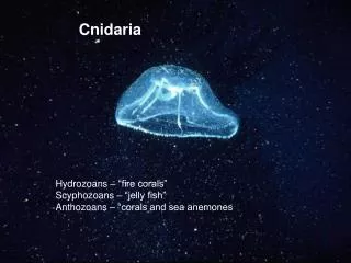
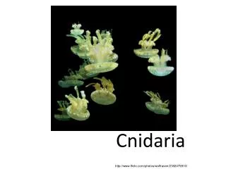
Scyphozoa • True jellyfish ~200 species • Mainly medusoid morphology
Fig. 1. Life cycle of the moon jellyfish Aurelia aurita. A Mature female medusa (30 cm in diameter) carrying planula larvae (red arrow) in brood pouches in the oral arms. B released, free-swimming planulae (0.2-0.3 mm). C Polyp (1-3mm). D Beginning strobilation. E Advanced strobilation. F Young Ephyra (3-5mm). G Ephyra, 4 weeks after release (8-10mm). The Moon Jelly Life Cycle!
Scyphozoan Sensory Structures • Sensory organs include: • Statocysts – balance organ • Ocelli – light receptor • Statocysts and ocelli are contained within structures called rhopalia • Cubozoans also have highly advanced sensory structures (lensed eyes)
CLEAN UP • Thoroughly wash all dissection tools and trays • Dispose of animal remains in biohazard container in the fume hood • Rinse slides & toss coverslips in sharps container • Make sure your scopes are clean and turned off • Present your worksheet
Feeding Anemone Feeding Video Anemone Feeding on Jellyfish Hydra Feeding Video Jellyfish Feeding