Download
1 / 14
140 likes | 353 Views
Connective Tissue Proper Cartilage Bone. Connective Tissue Proper: Loose Connective Tissue Dense Connective Tissue Adipose Tissue. Loose Connective Tissue: Locations: -Space between skin and internal body parts -adventitia of blood vessels -surrounds gland parenchyma (epi)

E N D
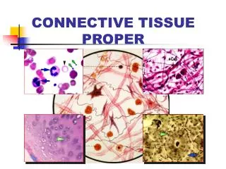
Connective Tissue Proper Cartilage Bone
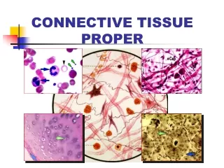
Connective Tissue Proper: Loose Connective Tissue Dense Connective Tissue Adipose Tissue
Loose Connective Tissue: Locations: -Space between skin and internal body parts -adventitia of blood vessels -surrounds gland parenchyma (epi) -lamina propria of GI tract
Loose Connective Tissue contains: -ground substance -fibroblasts -adipose cells -macrophages -mast cells -undifferentiated cells -collagen, elastic and reticular fibers
Dense Connective Tissue: Dense Irregular CT Dense Regular CT Dense Regular Collagenous CT Dense Regular Elastic CT Reticular Tissue Adipose Tissue
Dense Connective Tissue: • -same components as LCT • -fewer cells, more fibers • Dense Irregular CT - fiber arrangement is irregular • dermis of skin, nerve sheaths, capsules of spleen, • Testes, kindney, lymph nodes
Dense Regular Collagenous CT – coarsely packed collagen bundles densely packed - parrallel or sheet like orientation • little space for cells and ground substance • Thin, sheet-like fibroblasts between collagen bundles • Tendons and ligaments
Dense regular elastic connective tissue • Around large blood vessels • In the vertebral column • Has course branching elastic fibers • Few collagen fibers • Elastic fibers are arranged parallel to each other, forming thin sheets of fenestrated membranes
Elastic fibers • Found in LCT and as course bundles and sheets in dense regular elastic connective tissue • Made by fibroblasts of CT and by smooth muscle cells in blood vessels • Fibers contain the protein elastin (lots of glycine, proline, aa’s desmosine, isodesmosine cross-link elastin molecules.
Elastic fibers • Fibers contain the protein elastin (lots of glycine, proline, aa’s desmosine, isodesmosine cross-link elastin molecules and impart elasticity (can stretch) • Elastin in fiber core • Sheath of microfibrils (10 nm), a glycoprotein
Reticular Tissue • Framework (scaffolding) for liver, adipose tissue, bone marrow, lymph nodes, spleen etc. • Fibroblasts make type II collagen
Adipose Tissue • White (unilocular) • single lipid droplet, capillaries go into ct septa between fat cells. • Receptors for insulin, growth hormone, norepinephrine, glucocorticoids • Subcutaneous, in masses around the body
Adipose Tissue • Brown (multilocular) • Multiple droplets • Many mitochondria with cytochromes, lots of bv near the adipocytes • In human infants around the neck and interscapular region (?around eyes?)
Formation of adipocytes • Mesenchyme, may be a specific adipocyte precursor cell • Early Fetal: epitheloid precursors • Late fetal: secondary fat formation from fusiform precursor cells in ct.