Download
1 / 46
460 likes | 518 Views
Learn about the characteristics and classification of connective tissue proper, including loose connective tissue. Explore the different cell types, fibers, and ground substance present in this tissue.

E N D
Epithelium Connective tissue Pay attention to the differences b/w Epi & CT
一、CHARACTERISTICS: • Small number of cell large amount of intercellular matrix • Cell: separate and no polarity • Intercellular matrix: fibers + ground substance + tissue fluid • Filled with B.V., L.V., & N. • Function: support, connect, nourish, defence, and repair etc.
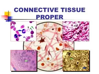
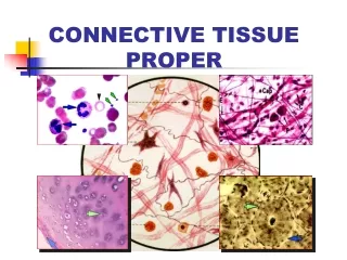
二、CLASSIFICATION • Connective tissue proper: Loose C.T. Dense C.T. Adipose tissue Reticular tissue • Cartilage and bone • Blood and Lymph
三、LOOSE CONNECTIVE TISSUE I. Characteristics: • Small number of cell & large number of cell category • small number of fibers & great amount of ground substance • Sponge-like structure (areolar tissue) • Distributed b/w cells, tissues and organs.
II. Cells • Fixed cells: fibroblasts fat cells undifferentiated mesenchymal cells • Wandering cells: macrophages plasma cells mast cells leukocytes
F.b. 1、Fibroblasts Structure: Nu: Cytoplasm: EM: rich in rER & r., developed G.l.
F.C. F.b. * Fibrocytes: Function: Synthesize & secrete collagenous pro. & elastic pro. (to form collagenous f., elastic f. & reticular f. ); and ground substance (proteoglycan & glycoprotein).
Mac. F.b. Mac. 2、Macrophages(Histocytes) Structure: Nu.: Cytoplasm:
EM:a large number of Ly., pinosome & phagosomes, bundles of MT & MF
Ag p.s. L.y 残余体 Function: • Mobility chemotaxis chemotactic factor: complement C5a, bacterial products etc. • Phagocytize specific:depend on identify factors: Ab, C, Fibronectin etc (the receptors of these factors are on surface of the macrophge). non-specific:independently
Ag p.s. L.y. • participate in immune regulation capture process antigen present antigen presenting molecular:MHCII Ag-MHCIIco. macrophage surface lymphocytes immune reaction
secrete bioactive products chemotactic factor(for polymorphonuclear leukocytes) immunosuppressive factor leukotriene interleukin(IL) interferon(INF) tumorous necrosis factor(TNF) etc. Source: themonocytes in blood
3、Plasma cells • Structure: Nu.:eccentrically,heterochromatin in wheel shape Cytoplasm: strong basophilic w/ lightly-stained area near Nu. P.C.
EM:rich in rER & r., developed Gl. • Source: B lmphocytes • Function: • synthesize immunoglobulin & cellular factors • participate in humoral immunity
4、Mast cells • Location & Structure: Nu.: Cytoplasm: basophilic granules, metachromatism M.C. B.V. Toluiding blue staining
EM: a great number of granules w/ crystals, containing: heparin, histamine, leukotriene, slow-reacting substance, eosinophil chemotactic factors (ECF-A)
Ag1 Ag2 R Ab-R degranulation plasma cell Ab(Ig E) • Function: • allergic reactions Ag1 plasma cell mast cell Ab(Ig E) R-Ab Ag2 degranulation • anticoagulation • attract eosinophil mast cell
. Ag1 Ab . Plasma cell . . . . . . . . . . . . . . . . . . . . . . . . . . . . . . . . . . . . . . . . . . . . . . . . . . . . . . . . . . . . . . . . . . . . . . . . Ag2 . . . . . . . . . . . . . . . . . . . . . . . . . . . . . . . . . . R-Ab Mast cell
Osmic acid staining HE staining B.V. N b 5、Fat cells (Adipose cells) • Shape: Nu.: Cytoplasm: • Function: synthesize & store lipid
6、Undifferentiated cells • Structure: similar to fibroblasts, smaller • Function: differentiate into various cell-types in C.T. during injury repairing e.g.:
L N 7、Leukocytes • Including: neutrophils, eosinophils, lymphocytes & monocytes • Function: defence involve in allergic reaction (see blood chapter)
III. Fibers: • Collagenous fibers (white f.) • Elastic fibers (yellow f.) • Reticular fibers (argyrophilic f.)
1、Collagenous fibers • Structure: LM: EM: fibrils:have periodic cross-bandings atintervals of 64nm fibrils
Collagen fibrils Collagen fibers
type I type III • Chemicals: collagen type I collagen type III Immunohistochemistry staining
Fibroblast ① ② Synthesize: In rER / GL: → Polypeptide αchains ① → hydroxylated αchains ② → Procollagen ③ In Extracellular space: → Tropocollagen ④ → fibrils ⑤ → collagenous f. ⑥ ③ ④ 280x1.4nm ⑤ ⑥
2、Elastic fibers Structure: LM: EM: elastin + microfibrils (no bandings) F.B.
3、Reticular fibers Sructure: LM:argyrophilia, PAS(+) EM: fibrils w/ periodic cross-bandings at intervals of 64nm, typeIII collagen covered carbohydrate
Reticular fibers in liver Reticular fibers in lymph node
IV.Ground substance • Jelly-like & amorphous substance; • Proteoglycan: in molecular sieve hyaluronic acid GAG(glycosaminoglycan) chondroitin sulfate keratin sulfate etc. protein: core protein & link protein glycoprotein:fibronectin (FN) laminin (LN) chondronectin (ChN);
Side chain subnit hyaluronic acid Collagenous fiber Ground substance Intercellular material
hyaluronic acid side chain subunit link pr. chondroitin sulfate keratin sulfate core pr. Link pr. molecular sieve: hyaluronic acid-- link pr.-- side chain subunit subunit: core protein + chondroitin sulfate & keratin sulfate tissue fluid: flowing through the sieve pores Core pr.
glycoprotein: • fibronectin (FN): produced by epithelial cells and fibroblasts play a role in events of identification, adhesion, migration and proliferation • laminin (LN) : located in B.M., produced by epi. cells, function: adhesion the epi.cells and B.M. • chondronectin (ChN): in cartilage tissue, fuction: a component of ground substances; adhesion chondrocyteS and colagen typeII
Function: • Tissue fluid nourishes the cells & tissues; • Molecular sieve acts as a barrier: to prevent the spread of bacteria & other microorganisms *haemolytic streptococci produce hyaluronidase & promote the invasion • Glycoproteins (FN/LN/ChN): identification /adherence/migration/proliferation etc.
四、DENSE CONNECTIVE TISSUE Characteristics: • Small amount of cells & ground substance, • large number of fibers • Fibers: wide, arranged densely • Function: connect & support
tendons dermis Lig. nuchae Category: Dense regular C.T. (tendons, cornea, ligment): parallel c.f. & tendon cells Dense irregular C.T. (dermis, sclera): collagenous fibers network & fibroblasts Elastic tissue (Lig. Nuchae, Lig. flavum & Large artery ): elastic fibers mainly Large artery
五、ADIPOSE TISSUE • Structure: Loose C.T.+ fat cells (in large aggregations) HE: / Osmic acid: • Function: energy storage, shock absorber, insulating layer
六、RETICULAR TISSUE cells Structure: • Reticular cells + reticular f. + G.S. • Reticular cell: stellate, pale nucleus, obvious nucleoli, processes (rich in rER) Function: architectural framework of lymphatic & hemato- poietic tissues Lymph node fibers