Connective Tissue and Cartilage
250 likes | 516 Views
Connective Tissue and Cartilage. 2007. Connective Tissue. Maintains the structural integrity of the body Functions of Connective Tissue provide structural support compartmentalize and encapsulate physical protection defense against infection storage of fat.

Connective Tissue and Cartilage
E N D
Presentation Transcript
Connective Tissue Maintains the structural integrity of the body Functions of Connective Tissue • provide structural support • compartmentalize and encapsulate • physical protection • defense against infection • storage of fat
Constituents of Connective Tissue • Cells – fibroblasts, adipocytes, immune cells • Extracellular matrix – consists of protein fibers embedded in a polysaccharide gel
Loose Connective Tissue of Mesentery Elastic fiber Fibroblast nucleus Collagen fiber
Principle Components of Extracellular Matrix • Ground substance • Composed of proteoglycans and glycosaminoglycans (GAGs) that are very hydrophilic • Forms a flexible gel through which metabolites diffuse • Protein fibers- provide tensile strength and elasticity • Collagen fibers – main fiber in connective tissue, forms bundles • Elastin fibers with microfibrils surrounding • Reticular fibers (Type III collagen), form mesh • Structural glycoproteins – cell adhesion molecules that mediate ECM and cell interactions • Examples: laminin, fibronectin, integrin
Reticular Connective Tissue Reticular fiber
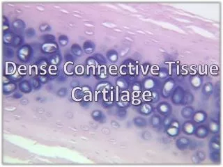
Cartilage Specialized form of connective tissue in which “fluid” ground substance is replaced by a solid substance that gives an added firmness to the tissue. chondroblast – cells of cartilage that are dividing and not fully differentiated, not surrounded by matrix. chondrocyte – cells of cartilage that are surrounded by matrix lacunae – spaces in the matrix where chondrocytes reside matrix – ground substance of cartilage perichondrium – region around outer edge of cartilage where developing chondroblasts reside
Growth of Cartilage Cartilage depends on diffusion (no blood vessels) for metabolite exchange between chondrocytes and surrounding tissues through the matrix – this limits the thickness of cartilage growth.
Hyaline Cartilage • Most abundant type of cartilage • Type II collagen fibers and matrix surround lacunae filled with chondrocytes. • Matrix smooth & uniform between lacunae. • Provides a low-friction surface • Location: nasal septum, larynx, tracheal rings, articular surfaces of bones
Elastic Cartilage • For strength and flexibility • Looks like hyaline cartilage and contains collagen • Also contains elastic fibers running in all directions • Location: external ear, walls of epiglottis, walls of Eustachian canals.
Elastic Cartilage – Low Power Elastic fiber
Fibrocartilage • Resists shearing forces • Has characteristics of both hyaline cartilage and dense regular connective tissue. • No perichondrium. • Location: intervertebral disks, pubic symphysis.