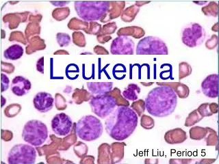
5.Types of Leukemia
770 likes | 840 Views
slideserveslideserve slideserveslideserveslideserve slideserveslideserveslideserve slideserveslideserveslideserve slideserveslideserveslideserve slideserveslideserveslideserve slideserveslideserveslideserve slideserveslideserveslideserve slideserveslideserveslideserve slideserve

5.Types of Leukemia
E N D
Presentation Transcript
What is the lymphatic system? • The lymphatic system is a network of tissues, vessels and organs that work together to move a colorless, watery fluid called lymph back into your circulatory system (your bloodstream). • Some 20 liters of plasma flow through your body’s arteries and smaller arteriole blood vessels and capillaries every day. • About 3 liters seep through the capillaries and into your body’s tissues. The lymphatic system collects this excess fluid, now called lymph, from tissues in your body and moves it along until it's ultimately returned to your bloodstream.
Function of lymphatic system • Maintains fluid levels in your body • Protects your body against foreign invaders • Transports and removes waste products and abnormal cells from the lymph
Parts of the lymphatic system • Lymph • Lymphatic vessels • Lymph nodes • Lymphatic organs • Primary lymphoid organs • Bone marrow (cells) • Thymus (T lymphocyte ) • Secondary lymphoid organs • Spleen • Lymph nodes
Causes of swollen lymph nodes • Infections • Strep throat – Measles - Ear infections Infected (abscessed) tooth – Mononucleosis - Skin or wound infections, such as cellulitis - Human immunodeficiency virus (HIV) - Tuberculosis- syphilis - Toxoplasmosis • Immune system disorders • Lupus - Rheumatoid arthritis • Cancers • Lymphoma - Leukemia • Other cancers (metastasized Cancer to lymph nodes)
Diagnosis • To diagnose what might be causing your swollen lymph nodes, your doctor may need: • Medical history. • Your doctor will want to know when and how your swollen lymph nodes developed and if you have any other signs or symptoms. • A physical exam. • Your doctor will also want to check lymph nodes near the surface of your skin for size, tenderness, warmth and texture. The site of your swollen lymph nodes and your other signs and symptoms will offer clues to the underlying cause. • Blood tests. • Certain blood tests may help confirm or exclude any suspected underlying conditions. The specific tests will depend on the suspected cause, but most likely will include a complete blood count (CBC). This test helps evaluate your overall health and detect a range of disorders, including infections and leukemia. • Imaging studies. • A chest X-ray or computerized tomography (CT) scan of the affected area may help determine potential sources of infection or find tumors. • Lymph node biopsy. • Your doctor may have you undergo a biopsy to secure the diagnosis. He or she will remove a sample from a lymph node or even an entire lymph node for microscopic examination.
Erythroblastic island Lipocyte Sinus Lipocyte Platelets Megakaryocyte Artery Central vein Sinus Endothelial cells
FETUS ADULT Yolk sac Axial skeleton Hematopoiesis Liver and spleen Distal long bones 01 2 3 4 5 6 7 891020 30405060 Month Year
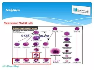
Hematopoiesis types • Intramedullary hematopoiesis • occur in the bone marrow • Extramedullary hematopoiesis • Occur outside the bone marrow • in the liver and spleen • the liver and spleen become enlarged
Posterior iliac crests Aspiration needle Stylet Hub Guard Skin Marrow Biopsy needle Hip bone Aspiration Biobsy
Cellularity • Cellularity is a hematopoietic cell/fat cell ratio • Normal cellularity = 1 • Cellularity > 1 refer as “Marrow Hyperplasia” • Cellularity < 1 refer as “Marrow Hypoplasia” • The mostly evaluated from biopsy specimen%)
Differential cell count • Granulocytic series (65%) (M) • Erythrocytic series (20%) (E) • Lymphocyte (10%) • Others (5%) • Myeloid:Erythroid ratio • (M:E ratio) = 2:1- 4:1 (300 to 500 cells are scanned) • presence of abnormal or tumor cells • The marrow is observed for abnormalities in the structure • (e.g., necrosis, fibrosis)
Iron accumulation • Storage iron is “hemosiderin” • It contained by nucleated erythroid cell • Prussian blue is the mostly stain for marrow iron storage • iron stores is benefit for evaluation of anemia
Spleen Function • Hematopoietic function • Can produce white blood cells, red blood cells, and platelets if necessary • Reservoir function • One-third of platelets and granulocytes are stored in the spleen • Filtration function • Aging red blood cells are destroyed; spleen removes inclusion from red blood cells; if red blood cell membrane is less deformable or antibody- coated, spleen presents a hostile environment leading to production of spherocytes • Immunologic function • Opsonizing antibodies are produced, trapping and processing antigens from encapsulated organs
Leukemia diagnosis • Complete blood picture (CBC) • Blood film • Bone marrow aspiration • Bone marrow biopsy • Immunophenotyping • Immunocytochemistry • Flow cytometry • PCR for genetic mutation
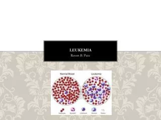
Leukemia Is a cancer that starts in blood-forming tissue, such as the bone marrow, and causes large numbers of abnormal or immatures blood cells to be produced and enter the bloodstream. • Chronic Leukemia A slowly progressing leukemia, and causes large numbers of abnormal mature or semi-mature WBCs to be produced and enter the blood stream • Acute Leukemia A fast progressing leukemia, and causes large numbers of abnormal immature WBCs to be produced and enter the blood stream • 20% of more blasts in the peripheral blood or bone marrow OR • The presence of a specific leukemia gene mutation.
Hematopoietic Stem cell Myeloid progenitor Lymphoid progenitor
Types of Leukemia • Lymphocytic Leukemia • Myeloid Leukemia • (Lymphoblastic) Leukemia • (Myelogenous) Leukemia (LL) (ML) Lymphocyte
Types of Leukemia • Lymphocytic Leukemia • Myeloid Leukemia • (lymphoblastic) Leukemia • (Myelogenous) Leukemia Acute Chronic Acute Chronic (AML) (CML) (ALL) (CLL)
Leukemoid Reaction • Defenition is typically a a physiological response to an underlying medical issue , involves having an elevated white blood cell count and or presencec of immature cells • Significance This term is used to differentiate between the leucocytosis and leukemia. • Characteristic • Toxic granulations. • Toxic vacuolization. • Presence of Dohle bodies. • No genetic defect