Download
1 / 46
460 likes | 479 Views
Anatomy of the Female Reproductive System. Liu Wei Department of Ob & Gy Ren Ji hospital. Introduction. Pelvis Pelvic floor External genitalia Internal genitalia Vessel and nerve and lymph Adjacent organs. Pelvis. Pelvis. Bony pelvis. os ilium 髂骨. os sacrum 骶骨. os coccyx 尾骨.

E N D
Anatomy of the Female Reproductive System Liu Wei Department of Ob & Gy Ren Ji hospital
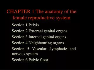
Introduction • Pelvis • Pelvic floor • External genitalia • Internal genitalia • Vessel and nerve and lymph • Adjacent organs
Pelvis • Bony pelvis os ilium 髂骨 os sacrum 骶骨 os coccyx 尾骨 os pubis 耻骨 os ischium 坐骨
Pelvis • Joints Sacro-iliac joint 骶髂关节 Sacro-coccygeal joint 骶尾关节 Symphysis pubica 耻骨联合
Pelvis • Ligaments • Sacrospinous ligament 骶棘韧带 Extend from the lateral border of the sacrum and coccyx to the spine of the ischium • Sacrotuberous ligament 骶结节韧带 Extend from the posterior aspect of the lower 3 sacral vertebrae to the ischial tuberosity
Pelvis • Pelvic divisions (iliopectineal line 髂耻线) • False pelvis (pelvis major) • Ture pelvis (pelvis minor) Bony birth canal the Pelvic inlet, the pelvic out let and the pelvic cavity
Pelvis • Types of pelvis • The gynecoid type (女型骨盆) round, slightly ovoid or elliptical inlet. adequate sacrosciatic notch. wide interspinous diameters(≥10cm). 52%-58.9% • The platypelloid type (扁平型骨盆) distinct oval inlet. very wide subpubic arch. 23.2%-29%
Pelvis • The anthropoid type (类人猿型骨盆) long, narrow, oval inlet. extended and narrow anterior and posterior segment. wide sacrosciatic notch. long , narrow sacrum. Straight side walls. • The android type (男型骨盆)
Pelvic floor • The tissues closing down the pelvic outlet (muscles and fasciae) suspend and support the pelvic organs, such as uterus and bladder and rectum • Posterior part (urogenital triangle) urethra and vagina pass through • Anterior part (anal triangle) rectum pass through
Pelvic floor • Tissues • Outer layer Bulbocavernosus muscle (球海绵体肌) Ischiocavernosus muscle (坐骨海绵体肌) Superficial transverse perineal muscle (会阴浅横肌) External anal sphincter (肛门外括约肌) • mid layer urogenital diaphragm泌尿生殖膈
Pelvic floor • Inner layer (pelvic diaphragm 盆膈) • the main support of the pelvic floor • formed by the levator ani and coccygenus muscles and covering fasciae. • Levator ani: pubococcygenus (耻尾肌), iliococcygenus (髂尾肌), puborectalis (坐尾肌)
Pelvic floor • Perineum general conception: the tissues closing down the pelvic outlet Clinical conception: the tissues between vagina and anus.
External genitalia mons pubis 阴阜 Labium majus 大阴唇 Clitotis 阴蒂 Labium minus 小阴唇 Urethral orifice 尿道口 Vaginal orifice 阴道口 Vaginal vestibule 阴道前庭 Fossa navicularis 舟状窝 Perineal body 会阴体 Anus 肛门
External genitalia • Labia majora The venous drainage is extensive and forms a plexus with numerous anastomoses. Vulva hematoma • Vaginal vestibule Bordered by the labia minora laterally, by the frenulum labiorum pudendal (阴唇系带) posteriorly, by the urethra and clitoris anteriorly, by the hymenal ring inferiorly.
Internal genitalia • Vagina • strong canal of muscle (7.5cm) extend from the uterus to the vestibule of the external genitalia. its long axis is almost parallel with that of the lower part of the sacrum. the anterior wall of the vagina is 1.5-2cm shorter than the posterior wall. • vaginal fornix the circular cul-de-sac formed around the cervix 4 regions: the anterior fornix, the posterior fornix and 2 lateral fornices.
Internal genitalia • Wall structure • mucosal layer (stratified squamous epithelium) • muscular layer (3 layer) • submucous area ( with a dense plexus of veins and lymphatics)
Internal genitalia • Uterus • Pear-shaped,thick-walled, muscular organ • Body and cervix: Babyhood 1:2, manhood 2:1 • Isthmus uteri connect the body to cervix, 1cm (non-pregnancy)
Internal genitalia • Layers of uterine wall • The serous layer (perimetrium) Thin and firmly adherent over the fundous and most of the body Uterovesical pouch of the peritoneum Rectouterine pouch of the peritoneum (pouch of Douglas) • The muscular layer Outer layer (longitudinal fibers) Inner layer (interlaced and various directions)
Internal genitalia • The mucous layer (endometrium) compact layer: response to hormone periodically, a single layer of ciliated columnar epithelium spongy layer: response to hormone periodically. contains many tubular glands funous: non-response to hormone periodically
Internal genitalia • Cervix • lower 1/3 of uterus. connects uterus to vagina via endocervical canal • External os: opening of endocervical canal to ectocervix • Internal os: indistinct upper limit of endocervical canal
Internal genitalia • Ligaments • Broad ligament • Round ligament • Cardinal ligament • Utero-sacral ligament
Internal genitalia • Oviduct • Anatomy • Interstitial portion: • Isthmic portion: narrow • Ampulla: wide and tortuous • Fimbria: funnel-shaped mouth
Internal genitalia • Layers of wall • Serous • Muscular: outer longitudinal and inner circular • Mucous: ciliated columnar epithelium, coarse longitudinal folds
Internal genitalia • Ovary • Anatomy • Paired organ, elliptic • The suspensory ligament of the ovary • The ovarian ligament
Internal genitalia • Structure of ovary • Covered by cuboid or low columnar epithelium • Consist of a cortex and a medulla • Cortex: oocytes in various stages of maturity. • Medulla: fibers, smooth muscle cells, blood vessel, nerves.
Vessel and nerve and lymph • Blood vessel • The ovarian artery • Orginated as branches of the abdominal aorta, (left: left renal artery). • Turn over the common iliac artery and ureter,descend into the pelvis. Enter into ovary through the mesovarium
Vessel and nerve and lymph • The uterine artery • a terminal branch of the hypogastric artery • Cross the ureter near the cervix (2cm) • Ascend along the lateral border of the uterus • uterine body branch and cervix-vagina branch
Vessel and nerve and lymph • Vaginal artery • Internal Pudendal artery
Vessel and nerve and lymph • Lymph • External genitalia • superfical inguinal gland (腹股沟浅淋巴结) • deep inguinal gland (腹股沟深淋巴结)
Vessel and nerve and lymph • Pelvic lymph • iliac lymph common iliac, internal iliac and external iliac • Anterior Sacral lymph • Lumbar lymph: abdominal aorta
Adjacent organs • Urethra • Urinary bladder • Ureter • Rectum • Vermiform appendix
Answer the questions • How many types of pelvis are there? What are they? • What is perineum? • What is vaginal vestibule? • How many ligaments of uterus are there? What are they? • How many ligaments of ovary are there? What are they? • What is uterine isthmus?