Download

1 / 12
0 likes | 50 Views
Understand bacterial shapes, sizes, and arrangements with clear visuals and examples. Covers cocci, bacilli, vibrio, spirochetes, and flagella types, along with microscopy techniques used in bacterial classification.<br>By Dr. Preeti Tyagi known as one of the best microbiology faculty in india.<br>ud83cudf10 www.turningbrain.in <br>ud83dudcde 91-8368648746<br>You can also watch her video lectures on YouTube as @dr.preetityagilectures874

E N D
Morphology of Bacteria -By Dr. Preeti Tyagi (MBBS,MD) Professor of Physiology VMMC & Sufdarjung Hospital New Delhi
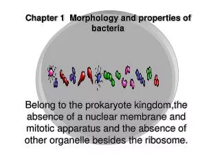
Introduction • Bacterial morphology refers to the size, shape, and arrangement of bacterial cells. It's one of the most fundamental aspects of microbiology and is crucial in identifying and classifying bacteria. • Understanding morphology helps in: • Diagnosing infections • Interpreting culture results • Studying bacterial evolution • The three key features of bacterial morphology include: • Shape (form of the cell) • Arrangement (how cells group after division) • Size (typically 0.2 – 2.0 µm in diameter)
Classification Based on Shape Vibrio (comma-shaped) Coccus (spherical) Spirochete (flexible spiral) E.g., Vibrio cholerae E.g., Streptococcus pneumoniae Bacillus (rod-shaped) Spirillum (rigid spiral) E.g., Treponema pallidum E.g., Spirillum minus E.g., Escherichia coli
Cocci Arrangement • Cocci can appear in various patterns based on their division: • Diplococci – in pairs (e.g., Neisseria gonorrhoeae) • Streptococci – in chains (e.g., Streptococcus pyogenes) • Staphylococci – in grape-like clusters (e.g., Staphylococcus aureus) • Tetrads – groups of four (e.g., Micrococcus luteus) • Sarcinae – cube of 8 cells (e.g., Sarcina ventriculi)
THANK-YOU • Full Physiology Course is available on Turning Brain App • by The Best Physiology Faculty in india, Dr. Preeti Tyagi • For More Info. +91-8368648746 www.turningbrain.in https://youtube.com/@dr.preetityagilectures874?si=YfbJEi66jOE0hjzu APP LINKS Android: https://play.google.com/store/apps/details id=tbrain.in.medical.eduapp&hl=en_IN IOS: https://apps.apple.com/in/app/turning-brain-dr-preeti-tyagi/id6502828714
Bacilli Arrangement • Rod-shaped bacteria (bacilli) have fewer arrangement types: • Single bacilli – e.g., Mycobacterium tuberculosis • Diplobacilli – two rods (e.g., Coxiella burnetii) • Streptobacilli – chains (e.g., Streptobacillus moniliformis) • Coccobacilli – short rods resembling cocci (e.g., Haemophilus influenzae)
Other Shaped Bacteria • Some bacteria have unique or irregular shapes: • Vibrio – curved rod, comma-shaped • Spirilla – rigid spiral with flagella • Spirochetes – flexible spirals, motile via axial filaments • Pleomorphic – variable shape due to lack of rigid cell wall (e.g., Mycoplasma)
Size of Bacteria • Bacteria’s are in different sizes: • Smallest: Mycoplasma (0.1 µm) • Largest: Epulopiscium fishelsoni (up to 750 µm) • Most pathogenic bacteria range from 0.5 to 5 µm • Size affects nutrient uptake, metabolism, and pathogenicity
Flagella and Motility • Flagella help in motility and classification: • Monotrichous – one flagellum (e.g., Vibrio cholerae) • Lophotrichous – multiple flagella at one end • Amphitrichous – flagella at both ends • Peritrichous – flagella all over the surface (e.g., E. coli)
Microscopy for Morphology • Bacterial morphology can be observed using: • Light Microscopy – Basic observation with staining • Gram Staining – Differentiates Gram-positive and Gram-negative • Electron Microscopy – Detailed 3D surface and internal structures
THANK-YOU • Full Physiology Course is available on Turning Brain App • by The Best Physiology Faculty in india, Dr. Preeti Tyagi • For More Info. +91-8368648746 www.turningbrain.in https://youtube.com/@dr.preetityagilectures874?si=YfbJEi66jOE0hjzu APP LINKS Android: https://play.google.com/store/apps/details id=tbrain.in.medical.eduapp&hl=en_IN IOS: https://apps.apple.com/in/app/turning-brain-dr-preeti-tyagi/id6502828714