Download
1 / 20
200 likes | 231 Views
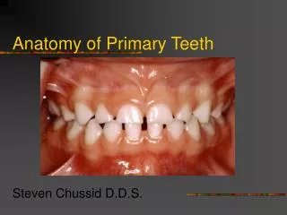
15.6.2015. Development of Teeth. Dr. Archana Rani Associate Professor Department of Anatomy KGMU UP, Lucknow. Introduction. Teeth are formed in relation to the alveolar process. Epithelial thickening: Dental lamina

E N D
15.6.2015 Development of Teeth Dr. Archana Rani Associate Professor Department of Anatomy KGMU UP, Lucknow
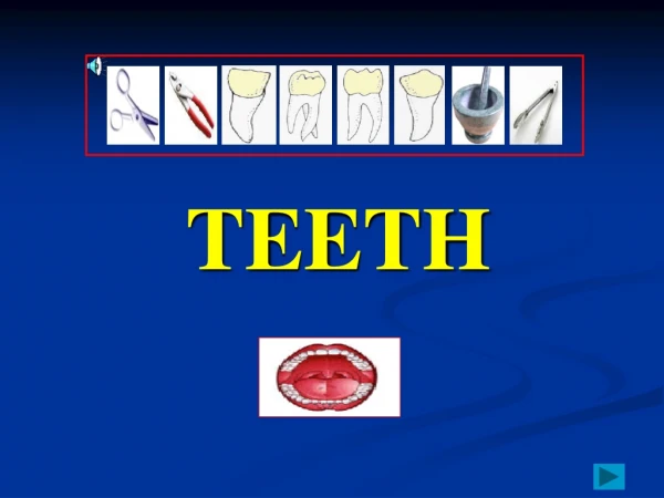
Introduction • Teeth are formed in relation to the alveolar process. • Epithelial thickening: Dental lamina • Enamel organs: Series of 10 localthickenings on dental lamina in each alveolar process. • Each thickening forms one milk tooth.
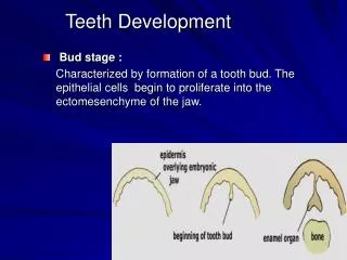
Bud stage : Characterized by formation of a tooth bud. The epithelial cells begin to proliferate into the ectomesenchyme of the jaw. Stages in the formation of enamel organ
Cap stage : • Formation of dental papilla. • The enamel organ & dental papilla forms the tooth germ. • Formation of ameloblasts. • Formation of odontoblasts.
Bell stage: The cells on the periphery of the enamel organ separate into three important layers: • Cuboidal cells on the periphery of the dental organ form the outer enamel epithelium. • The cells of the enamel organ adjacent to the dental papilla form the inner enamel epithelium. • The cells between the inner enamel epithelium and the stellate reticulum form a layer known as the stratum intermedium. The dental laminabegin to disintegrates, leaving the developing teeth completely separated from theepithelium of the oral cavity.
Crown stage : • Mineralization of hard tissues occur. • The inner enamel epithelial cells change in shape from cuboidal to columnar. The nuclei of these cells move closer to the stratum intermedium and away from the dental papilla. • The adjacent layer of cells in the dental papilla suddenly increases in size and differentiates into odontoblasts, which form dentin. • The inner enamel epithelium and the formation of odontoblasts continue from the tips of the cusps.
Crown stage (contd….): 5. The odontoblasts secrete an organic matrix into their immediate surrounding and form the dentin. 6. After dentin formation begins, the cells of the inner enamel epithelium secrete an organic matrix against the dentin. This matrix immediately mineralizes and becomes the tooth's enamel. 7. Outside the dentin are ameloblasts, which are cells that continue the process of enamel formation; therefore, enamel formation moves outwards, adding new material to the outer surface of the developing tooth.
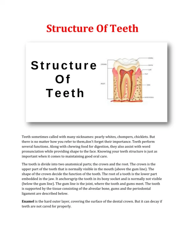
Structural components of teeth • Enamel: is a hard outer layer consisting primarily of calcium and phosphate in the form of hydroxyapatite. • Dentin: is the inner layer, the bulk of the tooth. • Pulp: is the core, containing nerves and blood vessels. • Cementum: is the thin layer around the root; a bone-like material which connects the teeth to the jaw.
Types of teeth according to their attachments • Acrodont:On the alveolar surface of the jaw. each tooth resides on the occlusional surface of the jaws in a very shallow socket. Replacement teeth arise adjacent to the active teeth (e.g. in snakes). • Thecodont : Teeth may be attached in sockets (replace within the same socket reptiles). with sockets (e.g. in mammals, crocodiles, dinosaurs). 3. Pleurodont: Attachment on the inner side of the jaws (e.g. in lizards). Teeth is continuously replaced.
Parts of a developing tooth An erupting temporary tooth
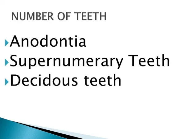
Anomalies of Teeth • Anodentia • Supernumerary teeth • Gemination • Malocclusion • Precocious eruption of teeth • Delayed eruption of teeth • Improper formation of enamel or dentine • Abnormal location of teeth e.g. in ovary or in hypophysis cerebri
REFERENCES 1. Langman’s Medical Embryology, 11th Edition. 3. I.B. Singh. Human Embryology, 10th Edition.
MCQs • The cells of the enamel organ that line the papilla are called: a) Ameloblasts b) Odontoblasts c) Cementoblasts d) Dental cuticle
MCQs 2. Mesenchymal cells which cover dentine are called: a) Ameloblasts b) Odontoblasts c) Cementoblasts d) Dental cuticle
MCQs 3. Temporary canines erupt at: a) 6-9 months b) 8-10 months c) 16-20 months d) 24-36 months
MCQs 4. The epithelium overlying the alveolar process become thickened to form: a) Dental lamina b) Dental papilla c) Dental cuticle d) Periodontal ligament
MCQs 5. Absence of ameloblasts is a feature of: a) Crown b) Enamel c) Dentine d) Root