Download
1 / 30
910 likes | 4k Views
Anatomy of Primary Teeth. Steven Chussid D.D.S. Lecture Overview. Primary Dentition General Morphological considerations Implications of Primary tooth morphology. Primary Dentition. 20 primary teeth as compared to 32 permanent teeth No premolars in the primary dentition

E N D
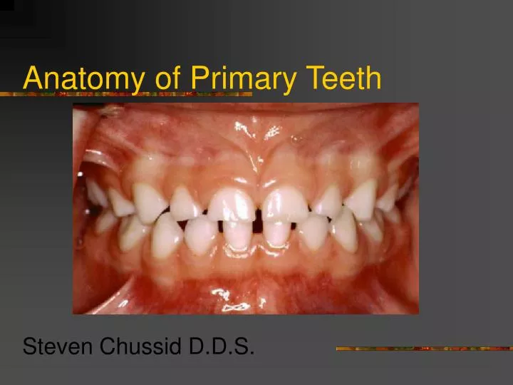
Anatomy of Primary Teeth Steven Chussid D.D.S.
Lecture Overview • Primary Dentition • General Morphological considerations • Implications of Primary tooth morphology
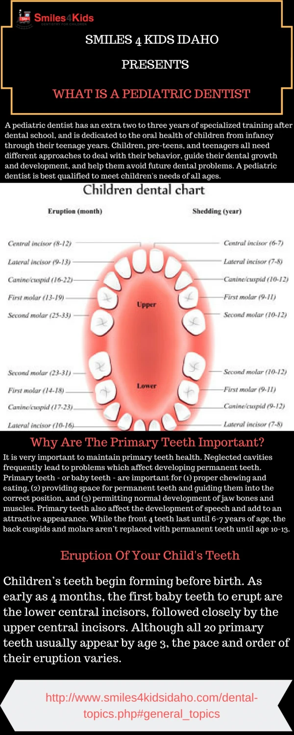
Primary Dentition • 20 primary teeth as compared to 32 permanent teeth • No premolars in the primary dentition • The primary molars are replaced by the premolars • The permanent molars erupt distal to the primary second molars
Eruption Patterns of Primary Teeth • Sequence more important than timing • Symmetrical pattern • Mandibular teeth erupt first
Approximate Eruption Schedule “7+4” Guideline
11 Months = 4 Erupted Teeth • 7 months + 4 = 11 months • 0 teeth + 4 = 4 teeth
15 Months = 8 Erupted Teeth • 11 months + 4 = 15 months • 4 teeth + 4 = 8 teeth
19 Months = 12 Erupted Teeth • 15 months + 4 = 19 months • 8 teeth + 4 = 12 teeth
23 Months = 16 Erupted Teeth • 19 months + 4 = 23 months • 12 teeth + 4 = 16 teeth
27 Months = 20 Erupted Teeth • 23 months + 4 = 27 months • 16 teeth + 4 = 20 teeth
General Morphologic considerations • Crown • Pulp • Root
Crown of Primary Teeth • Shorter • Narrower occlusal table • Constricted in the cervical portion • Thinner enamel and dentin layers • Enamel rods in the cervical area directed occlusally • Broad and flat contacts • Color is usually lighter
Crowns of Primary Teeth • Prominent mesio-buccal cervical bulge seen in primary molars • Incisors have no developmental grooves or mammelons
Primary Crown Anatomy • Mandibular Central Incisors- • Symmetrically flat when viewed from buccal • Crown about 1/3 length of root • Cingulum present on lingual surface • Mandibular Lateral Incisor • Similar form to central • Usually longer • Incisal edge slopes toward distal and DI angle more rounded
Primary Crown Anatomy • Maxillary Central Incisor • Only tooth that has a greater mesiodistal width than height • Prominent cingulum • Incisal edge straight • Maxillary Lateral Incisor • Similar form to cental • Smaller and DI angle rounded
Primary Crown Anatomy • Maxillary Canine • Crown constricted at cervical region • Well developed, sharp cusp • Root is long, more than 2X crown • Mandibular Canine • Similar form to maxillary • Crown shorter and narrower labiolingually
Primary Crown Anatomy • Maxillary First Molar • Unique appearance • Three cusps-MB, DB and Lingual • Prominent MB cervical bulge • Mandibular First Molar • Also unique in appearance • Four cusps-MB, DB, ML and DL • Prominent MB cervical bulge • Transverse ridge
Primary Crown Anatomy • Maxillary 2nd Molar • Resembles permanent maxillary first molar but smaller • Mandibular 2nd Molar- • Resembles permanent mandibular first molar but smaller
Pulps of Primary Teeth • Relatively larger • Pulp horns are closer to the outer surface • Great variation in size and location • Mesial pulp horn is higher • Pulp chamber shallow • Form of the pulp follows the external anatomy • Usually a pulp horn under each cusp
Roots of Primary Teeth • Roots of anterior teeth are narrower mesio-distally • Posterior teeth have longer and more slender roots in relation to crown size • Molar roots flare more as they approach the apex • Apical foramina may be larger and accessory canals often larger and more numerous
Implications of Primary tooth morphology • The progress of caries is much faster in the primary dentition, so incipient lesions should be restored sooner than later! • Thinner enamel and dentin • Mesial pulp horn higher
Procedures in Primary Teeth • Restorative Dentistry • Enamel is thinner, therefore modifications are necessary in the cavity prep • Broad contacts need to be restored • Beware of the mesio-buccal pulp horn • May need to do SSC if both proximal surfaces involved • Preserve the buccal cervical ridge to obtain mechanical retention for SSC
Procedures in Primary Teeth • Surgical Procedures • Conical anterior roots facilitate easy removal • Flared roots of the molars - use caution as premolar buds are located between the roots • Pulp Therapy • Pulpotomy- beware of perforations • Pulpectomy • Difficult on molars due to tortuous and irregular pulp canals • Beware of tooth buds
Summary • Primary teeth have • Thinner enamel and dentin layers • Pulp horns closer to the outer surface • Mesial pulp horn much higher • Relatively larger pulps • Enamel rods direct slightly occlusally in the cervical area • Cervical area is constricted significantly • Roots flare as they approach the apex • More tortuous and irregular pulp canals