Download
1 / 7
190 likes | 782 Views
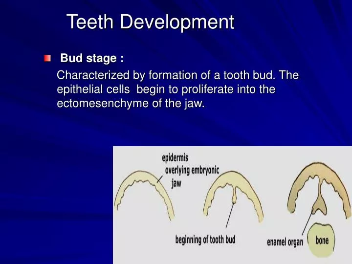
Teeth Development. Bud stage : Characterized by formation of a tooth bud. The epithelial cells begin to proliferate into the ectomesenchyme of the jaw. . Cap stage :

E N D
Teeth Development • Bud stage : Characterized by formation of a tooth bud. The epithelial cells begin to proliferate into the ectomesenchyme of the jaw.
Cap stage : The ectomesenchymal cells aggregated forming the dental papilla. At this point, the tooth bud grows around the ectomesenchymal aggregation, taking on the appearance of a cap, and becomes the enamel (or dental) organ. The enamel organ will produce enamel, the dental papilla will produce dentin and pulp, and the dental follicle will produce all the supporting structures of a tooth.
Bell stage : The cells on the periphery of the enamel organ separate into three important layers: • Cuboidal cells on the periphery of the dental organ and form the outer enamel epithelium. • The cells of the enamel organ adjacent to the dental papilla and form the inner enamel epithelium. • The cells between the inner enamel epithelium and the stellate reticulum and form a layer known as the stratum intermedium. The dental laminabegin to disintegrates, leaving the developing teeth completely separated from theepithelium of the oral cavity.
Crown stage : • During this stage mineralized hard tissues ocurred. • The inner enamel epithelial cells change in shape from cuboidal to columnar. The nuclei of these cells move closer to the stratum intermedium and away from the dental papilla. • The adjacent layer of cells in the dental papilla suddenly increases in size and differentiates into odontoblasts, which are the cells that form dentin.. • The inner enamel epithelium and the formation of odontoblasts continue from the tips of the cusps. • The odontoblasts secrete an organic matrix, into their immediate surrounding and forming the dentin . • After dentin formation begins, the cells of the inner enamel epithelium secrete an organic matrix against the dentin. This matrix immediately mineralizes and becomes the tooth's enamel. • Outside the dentin are ameloblasts, which are cells that continue the process of enamel formation; therefore, enamel formation moves outwards, adding new material to the outer surface of the developing tooth.
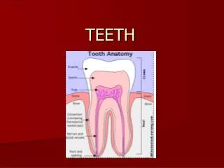
Structural components of teeth: • Enamel : is a hard outer layer consisting primarily of calcium and phosphate in the form ofhydroxyapatite. • Dentin : is the inner layer, the bulk of the tooth. • Pulp : is the core, containing nerves and blood vessels. • Cementum : is the thin layer around the root; abone-like material which connects the teeth to the jaw.
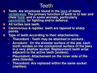
Type of teeth according to their attachments: • Acrodont: On the alveolar surface of the jaw. each tooth resides on the occlusional surface of the jaws in a very shallow socket. Replacement teeth arise adjacent to the active teeth (snakes). • Thecodont : Teeth may be attached in sockets . ( replace within the same socket reptiles). with sockets (mammals, crocodiles, dinosaurs ). • Pleurodont: Attachement on the inner side of the jaws (lizards). Teeth continuously replaced.