Download
1 / 105
1.11k likes | 1.88k Views
PEMERIKSAAN LABORATORIUM KELAINAN UROGENITAL. Efrida, dr., SpPK., MKes. Staf Pengajar Patologi Klinik FK UNAND SMF Patologi Klinik RS. Dr. M. Djamil Padang 7 Maret 2013.

E N D
PEMERIKSAAN LABORATORIUM KELAINAN UROGENITAL Efrida, dr., SpPK., MKes Staf Pengajar Patologi Klinik FK UNAND SMF Patologi Klinik RS. Dr. M. Djamil Padang 7 Maret 2013
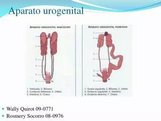
Urinalisis/Analisis Urin Memberi informasi: • Keadaan Ginjal dan saluran kemih • Faal hati • Saluran empedu • Pankreas • Korteks adrenal • Dll
Komponen Urin Normal • Air (95%) • Produk sisa terlarut: ureum, kreatinin, as. Urat • Elektrolit: Na, K, Cl, Ca, Fosfat • Hormon: setelah menjalankan fungsi • Komposisi lain: tergantung makanan/cairan/obat yang dikonsumsi
Tujuan Urinalisis • Menunjang diagnosis gangguan ginjal, traktus urinarius, dan penyakit sistemik yang mempengaruhi fungsi ginjal • Memantau perjalanan penyakit (misal: DM) • Memantau efektivitas pengobatan/komplikasi • Skrining dan pemantauan penyakit asimptomatik kongenital/herediter • Skrining drug abuse • General check up
Indikasi Permintaan Urinalisis • Gejala/riwayat penyakit ginjal/sal. Kemih • Gangguan endokrin • Ikterik • Terapi yg mempengaruhi fungsi ginjal • Kehamilan • Toksikologi/over dosis obat/narkoba • Abnormalitas genetik(gangguan metabolisme AA)
Tahap pemeriksaan: • Praanalitik a. persiapan pasien b. persiapan sampel teknik sampling yg baik wadah penampung bersih, kering, bermulut lebar. tes biakan urin wadah dan metode sampling harus steril
Pedoman NCCLS • Identifikasi sampel: nama, MR, alamat/ruang rawat, penggunaan pengawet • Specimen acceptability Urinalisis dilakukan dalam waktu < 2 jam setelah dikemihkan. Jika ditundarefrigerator Sampel tanpa label/identitastolak Hindari kontaminasi • Kontrol kualitas
c. Cara pengumpulan sampel - sering pengumpulan urin ketika berkemih suatu saat (urine sewaktu) - kateterisasirisiko infeksi - punksi suprapubik - clean catch/clean voided midstream
Urine sewaktu /random • Urine pagi • Urine postprandial / 2 jam pp • Urine 24 jam • Jenis sampel urine • Urine sewaktu Yang dikeluarkan pada satu waktu yg tidak ditentukan dengan khusus (u/ pem rutin, skrining, tanpa saran khusus) • Urine pagi Yang dikeluarkan kedua kali pada pagi hari setelah bangun tidur sebelum makan dan sebelum gerak badan( urine lebih pekat). (u/ pem sedimen, BJ, protein, kehamilan)
Urine 2 jam PP Yang dikeluarkan pertama kali 2 jam Setelah makan. (u/ pem glukosa) • Urine 24 jam u/ penetapan kuantitatif sesuatu zat dalam urine Perlu pengawet PATIENT GETS UP URINATES (AT 06.00) DISCARDED AT 06.00 THE OTHER DAY URINE PASSED DURING THE REST OF THE DAY
Pemeriksaan rutin • Urine baru • Jika terpaksa + pengawet urine • Jenis pengawet urine • Toluena • Thymol • Formaldehida • Asam sulfat pekat • Natriumkarbonat • Wadah urine • Besih dan kering • Bermulut lebar • Tutup rapat
URINE BARU ADUK ~ HOMOGEN • MAKROSKOPIK • WARNA • BAU • KEKERUHAN • KEASAMAN • BERAT JENIS • VOLUME SEDIMEN SUPERNATAN • KIMIA • ALBUMIN • GLUCOSE • UROBILIN • BILIRUBIN • KETOBODY • BENZIDIN • MIKROSKOPIK • ERITROSIT • LEUKOSIT • EPITEL • KRISTAL • CAST
1. MAKROSKOPIK URINE WARNA • KUNING MUDA NORMALUROKROM • KUNING TUA BILIRUBIN (?) • FOAM TEST (+) KUNING (JELAS) HAWKINSON/HARISON (-) / MERAGUKAN FOUCHET KOCOK (KUAT-KUAT) FOAM • MERAH (DARAH?) • SEDIMEN ERITROSIT : (+) = HEMATURI • (-) BENZIDIN TEST
KEKERUHAN (NORMAL : JERNIH) • KEMERAHAN DARAH SEDIMEN ? • (ERITROSIT) • BERKABUT BAKTERI (GRAM) • KERUH (ALKALIS / URINE NETRAL) - PUS - FOSFAT / KRISTAL KARBONAT • BERKURANG / HILANG • (FOSFAT/KRISTAL KARBONAT) • SPERMATOZOA + ASAM ASETAT (6%)
Color and clarity • Color : normally , pale to dark yellow (urochrome) Abnormal color : some drugs cause color changes 1. red urine: causes: hematuria hemoglobinuria myoglobinuria 2. yellow-brown or green-brown urine: bilirubin cause : obstructive jaundice
Microscopic Hematuria Urinary tract source Urethra or bladder Prostate Ureter or kidney Non-Urinary tract source Vagina Anus or rectum Pseudohematuria (non-hematuria related red urine) Myoglobinuria Hemoglobinuria PhenolphthaleinLaxatives Phenothiazines Porphyria Rifampin Pyridium Bilirubinuria Phenytoin Pyridium Red diaper syndrome Foods (Beets, Blackberries, Rhubarb) Red Urine
Red Urine • Causes of Asymptomatic Gross Hematuria by Incidence • Acute Cystitis (23%) • Bladder Cancer (17%) • Benign Prostatic Hyperplasia (12%) • Nephrolithiasis (10%) • Benign essential hematuria (10%) • Prostatitis (9%) • Renal cancer (6%) • Pyelonephritis (4%) • Prostate Cancer (3%) • Urethral stricture (2%)
Clarity: normally, clear Abnormal color: cloudy urine Causes: 1. crystals or nonpathologic salts phosphate, carbonate in alkaline urine (dissolve---add acetic acid) uric acid in acid urine (dissolve---warming to 60℃) 2. various cellular elements: leukocytes, RBCs, epithelial cells
Urine volume • The average adult : 1000ml to 2000ml/24h • Increase polyuria---more than 2000ml of urine in 24 hours 1. physiological states: water intake, some drugs, intravenous solutions 2. pathologic states: diabetes mellitus, diabetes insipidus
Urine volume • Decrease Oliguria---less than 400ml of urine in 24 hours Anuria---less than 100ml of urine in 24 hours 1. prerenal: hemorrhage, dehydration, congestive heart failure 2. postrenal: obstruction of the urinary tract (may be stones, carcinoma) 3. renal parenchymal disease: acute tubular necrosis, chronic renal failure
Specific gravity (SG) • Reflect the density of the urine • Range of 1.001 to 1.040 • Increase: Dehydration、Fever、VomitingDiarrheaDiabetes Mellitus and other causes of Glycosuria、Congestive Heart Failure、Syndrome Inappropriate ADH Secretion (SIADH) 、Adrenal Insufficiency failure (urine volume↓ and SG↑) • Decrease: diabetes insipidus (urine volume↑ and SG ↓)
BERAT JENIS • REFRACTOMETER • KEUNTUNGAN : • BAHAN SEDIKIT • MUDAH • KERUGIAN : • < AKURAT • HARUS DIKALIBRASI : • SUHU • GLUKOSA • PROTEIN - URINOMETER 1,000 KEUNTUNGAN : -> AKURAT KERUGIAN : -BAHAN BANYAK 1,020 KOREKSI 1,040 - CARIK CELUP
Urine PH • Normal PH The average is about 6 Range from 5~9 (depends on diet)/4,7-7,5 • Higher PH---alkaline urine 1.drugs: sodium bicarbonate 2.classic renal tubular acidosis 3.alkalosis (metabolic or respiratory) • Lower PH---acid urine 1.drugs: ammonium chloride 2. acidosis (metabolic or respiratory)
KEASAMAN (pH) (N. 4,7 – 7,5) RATA-RATA : 6,0 • KERTAS LAKMUS • BIRUMERAH= ASAM • BIRU = NORMAL MERAH = NORMAL • RED BIRU = BASA/ALKALIS METODE LAIN: CARIK CELUP
B A U • NORMAL BAU URINE (Bau ureum) KARENA ADANYA UREUM DLM URINE • ABNORMAL c/ BAU JENGKOL • INTOKSIKASI JENGKOL • + ALBUMINURIA • HEMATURIA • KRISTALURIA • BUAH-BUAHAN KETONURIA
2. MIKROSKOPIK URINE URINE SENTRIFUS 1500 RPM / 5 MNT TUTUP DGN COVER GLASS SEDIMEN TETESKAN SLIDE MIKROSKOP OBJECTIVE 40 X DAN 10 X OCULAR 10 X CONDENSOR PEMERIKSAAN ! ! ERITROSIT/ HIGH POWER LEUKOSIT / HIGH POWER SEDIMEN ORGANIK SILINDER / LOW POWER SEL EPITEL KRISTAL ANORGANIK SEDIMEN
LEUKOSIT NORMAL : 0 - 6 / LPB MORFOLOGI : • BAGIAN PINGGIRNYA TIDAK JELAS • UKURAN ; F+ 11 m • GRANULA (+) DLM SITOPLASMA E ERITROSIT NORMAL : 0 - 1 / LPB MORFOLOGI : • A.NORMAL (URINE BARU) : • - UKURAN, F + 7 m • - KEKUNINGAN • - PINGGIRNYA JELAS • CRENATED (BJ URINE TINGGI)
Pemeriksaan Kimiawi BENEDICT (PEMERIKSAAN GLUKOSA) • Prinsip: Dalam suasana alkali kuat, gula-gula (reduktor) akan mereduksi Cupri menjadi Cuprohidroksida (CuOH) yang berwarna kuning atau Cuprooksida (CuO) yang berwarna merah • Metode : Benedict (manual) • Reagen : CuSO4.5H2O …………………17,3 gram Na Carbonat Anhidrous ……… 100 gram Na Citrat ……………………... 173 gram Dilarutkan dalam 1 liter akuades