Download
1 / 33
330 likes | 775 Views
Neurogenic Bladder. Prepared by Dr. Abdullah Ghazi (R4) Supervised by Dr. Ali Binmahfooz 1/12/2010 KFSH&RC. Subject. Anatomy and physiology Classification of neurogenic LUTS. Evaluation. Management. Anatomy and Physiology. Bladder - Anatomy. Neuroanatomy of Voiding.

E N D
Neurogenic Bladder Prepared by Dr. Abdullah Ghazi (R4) Supervised by Dr. Ali Binmahfooz 1/12/2010 KFSH&RC
Subject • Anatomy and physiology • Classification of neurogenic LUTS. • Evaluation. • Management.
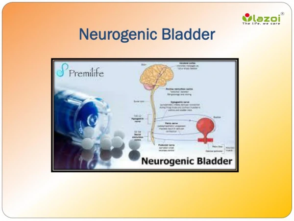
Neuroanatomy of Voiding • Frontal lobe • Micturition center • Sends inhibitory signals • Pons (Pontine Micturition Center) • Excitatory center • Coordinates urinary sphincters and the bladder • Spinal cord • Intermediary between upper and lower control
Peripheral Nervous System • Somatic (S2-S4) • Pudendal nerves • Excitatory to external sphincter • Parasympathetic (S2-S4) • Pelvic nerves • Excitatory to bladder, relaxes sphincter • Sympathetic (T10-L2) • Hypogastric nerves to pelvic ganglia • Inhibitory to bladder, excitatory to urethra
Normal Voiding • SNS primarily controls bladder and the IUS • Bladder increases capacity but not pressure • Internal urinary sphincter to remain tightly closed • Parasympathetic stimulation inhibited • Somatics (pudendal N) regulate • External urinary sphincter • Pelvic diaphragm • PNS • Immediately prior to PNS stimulation, SNS is suppressed • Stimulates detrusor to contract • Pudendal nerve is inhibited external sphincter opens facilitation of voluntary urination
Pathophysiology of Voiding • Brain lesion above pons destroys master control center • Stroke (35%) • Brain tumor (24%). • Hydrocephalus (22%). • CP (35%). • Mental retarted (50%) • Basal ganglia pathology (40%) • Result : • urge incontinence. • night incontinence. • coordinated sphincter
Pathophysiology of Voiding • Spinal cord. • Spinal cord lesion (95%). • Myelomeningocele (50%DSD). • Multiple Sclerosis (70%). • Result: • Detrusor hyperreflexia & spastic bladder. • Detrusor Sphincteric Dyssynergia. • Some: Areflexic bladder
Pathophysiology of Voiding • Lumbosacral spinal lesion • Spinal tumor. • Herniated disc (50%). • Lumbar laminectomy (50%). • Radical hysterectomy. • Pelvic trauma • Result – areflexic bladder
Pathophysiology of Voiding • Peripheral nerve injury • Diabetes (50-25%). • Polio. • Alcohol abuse • GBS.
History • General history • Specific history • Urinary history • Bowel history: • Sexual history • Neurological history
Examination • Sensation S2-S5 on both sides of the body • Reflexes • Anal sphincter tone
Investigation • Urinalysis • Blood chemistry • Voiding diary • Residual urine (UFM). • Quantification of urine loss by pad testing if appropriate • Urinary tract imaging studies • Urodynamic study.
Finding at Urodynamic • Filling phase • Hyposensitivity or hypersensitivity • Vegetative sensations • Low compliance • High capacity bladder • Detrusor overactivity, spontaneous or provoked • Sphincter acontractility.
Finding at Urodynamic • Voiding phase • Detrusor acontractility • DSD • Non-relaxing urethra • Non-relaxing bladder neck
GUIDELINES FOR URODYNAMICS AND URO-NEUROPHYSIOLOGY • Urodynamic investigation is necessary to document the dysfunction of the LUT (A). • The recording of a bladder diary is advisable (B). • Non-invasive testing is mandatory before invasive urodynamics is planned (A). • Video-urodynamics is the gold standard for invasive urodynamics in patients with NLUTD. If this is available, then a filling cystometry continuing into a pressure flow study should be performed (A). • A physiological filling rate and body-warm saline must be used (A). • Specific uro-neurophysiological tests are elective procedures (C).
Treatment Priority 1. Protection of the upper urinary tract 2. Improvement of urinary continence 3. Restoration of (parts of) the LUT function 4. Improvement of the patient’s quality of life.
Goal of Treatment • In patients with high detrusor pressure (detrusor overactivity, low detrusor compliance, DSD, other causes of bladder outlet obstruction). • Aim to conversion high-pressure bladder into a passive low-pressure reservoir despite the resulting residual urine.
Non-invasive Conservative Treatment • Assisted bladder emptying, Credé, Valsalva. • Lower urinary tract rehabilitation • Behavioural modification techniques • Pelvic floor muscle exercises • Pelvic floor electrostimulation • Biofeedback • Drug treatment • Anticholinergic agents • Phosphodiesterase inhibitors, desmopressin. • Cholinergic drugs (bethanechol chloride). • Alpha-blockers. • Increasing bladder outlet resistance (no puplish). • Electrical neuromodulation • External appliances
GUIDELINES FOR NON-INVASIVE CONSERVATIVE TREATMENT • The first aim of any therapy is the protection of the upper urinary tract. • The mainstay of treatment for overactive detrusor is anticholinergic drug therapy (A) • Lower urinary tract rehabilitation may be effective in selected cases. • Condom catheter or pads may reduce urinary incontinence to a socially acceptable situation. • Any method of assisted bladder emptying should be used with the greatest caution (A).
Minimal Invasive Treatment • Catheterization • Intravesical drug treatment • Intravesicalelectrostimulation • Botulinum toxin injections in the bladder • Bladder neck and urethral procedures • Botulinum toxin sphincter injection • Balloon dilatation • Sphincterotomy • Stents • Bladder neck incision • Increasing bladder outlet resistance
GUIDELINES FOR CATHETERIZATION • Intermittent catheterization is the standard treatment for patients who are unable to empty their bladder (A). • Patients should be well instructed in the technique and risks of IC. • Aseptic IC is the method of choice (B). • The catheter size should be 12-14 Fr (B). • The frequency of IC is 4-6 times per day (B). • The bladder volume should remain below 400 mL (B). • Indwelling transurethral and suprapubic catheterization should be used only exceptionally, under close control, and the catheter should be changed frequently. Silicone catheters are preferred and should be changed every 2-4 weeks, while (coated) latex catheters need to be changed every 1-2 weeks. (A).
GUIDELINES FOR MINIMAL INVASIVE TREATMENT • Botulinum toxin injection in the detrusor is the most effective minimally invasive treatment to reduce neurogenic detrusor overactivity (A). • Sphincterotomy is the standard treatment for DSD (A). • Bladder neck incision is effective in a fibrotic bladder neck (B).
Surgical Treatment • Urethral and bladder neck procedures • Urethral sling • Artificial urinary sphincter • Functional sphincter augmentation (gracilis m) • Bladder neck and urethra reconstruction (Extrophy) • Detrusormyectomy (auto-augmentation) • Denervation, deafferentation, neurostimulation, neuromodulation • Bladder covering by striated muscle (rectus m) • Bladder augmentation or substitution • Urinary diversion (continent diversion, incontinent diversion)
GUIDELINES FOR SURGICAL TREATMENT • Detrusor • Overactive • Detrusor myectomy is an acceptable option for the treatment of overactive bladder when more conservative approaches have failed. It is limited invasive and has minimal morbidity (B). • - Sacral rhizotomy with SARS in complete lesions and sacral neuromodulation in incomplete lesions are effective treatments in selected patients (B). • Bladder augmentation is an acceptable option for decreasing detrusor pressure whenever less invasive procedures have failed. For the treatment of a severely thick or fibrotic bladder wall, a bladder substitution might be considered (B).
GUIDELINES FOR SURGICAL TREATMENT • Detrusor • Underactive • SARS with rhizotomy and sacral neuromodulation are effective in selected patients (B). • Restoration of a functional bladder by covering with striated muscle is still experimental (4).
GUIDELINES FOR SURGICAL TREATMENT • Urethra • Overactive (DSD): like minimal invasive treatment • Underactive • The placement of a urethral sling is an established procedure (B). • The artificial urinary sphincter is very effective (B). • Transposition of the gracilis muscle is still experimental (Level of evidence: 4).
Referance • European Association of Urology 2010 M. Stöhrer, B. Blok, D. Castro-Diaz, E. Chartier-Kastler, G. Del Popolo, G. Kramer, J. Pannek, P. Radziszewski, J-J. Wyndaele