Download
1 / 20
240 likes | 507 Views
Bile canaliculi are connected to interlobular bile duct by means of short narrow channels called bile ductules or canal of Hering . They are lined by cuboidal epithelium. (have property of proliferation so called as hepatic stem cells)

E N D
Bile canaliculiare connected to interlobular bile duct by means of short narrow channels called bile ductules or canal of Hering. • They are lined by cuboidal epithelium. (have property of proliferation so called as hepatic stem cells) • The interlobular bile ducts form one of the three components of the portal triad. • Bile ducts are lined by simple columnar epithelium • They fuse with each other and form hepatic duct (Rt and Lt)
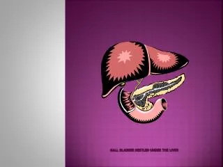
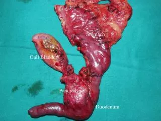
Biliary Tract & Gallbladder • The bile produced by the hepatocytes flows through the bile canaliculi, bile ductules, and bile ducts. • These structures gradually merge, forming a network that converges to form the hepatic duct. • The hepatic duct, after receiving the cystic duct from the gallbladder, continues to the duodenum as the common bile duct
The hepatic, cystic, and common bile ducts are lined with a mucous membrane having a simple columnar epithelium of cholangiocytes. • Has three coats • 1. mucosa • 2. muscularis • 3. adventitia/serosa
Mucosa consists of epithelium and lamina propria is thrown into numerous folds (prominent in empty gallbladder) • Simple columnar epithelium. Tall cells with oval basal nuclei. • Free surface of cells bear microvilli (presence of thin striated border) . • Lamina propriaconsists of loose connective tissue rich in elastic and collagen fibres. Has fenestrated capillaries, lymphocytes, venulesand plasma cells • devoid of glands except in neck region where simple tubuloacinar glands are present which secrete mucus • Submucosais absent
Muscularispropriabecomes thicker near the duodenum and finally, in the portion within the duodenal wall, forms a sphincter that regulates bile flow. • Smooth muscle fibres are present and collagen and elastic fibres in between. • Adventitia is present where gall bladder is attached to the liver other wise serosa is present.
The lining epithelial cells have prominent mitochondria, microvilli, and intercellular spaces, all indicative of active absorptive cells • The main function of the gallbladder is to store bile, concentrate it by absorbing its water, and release it when necessary into the digestive tract. • This process depends on an active sodium-transporting mechanism in the gallbladder's epithelium, with water absorption from bile an osmotic consequence of the sodium pump.
Contraction of the smooth muscle of the gallbladder is induced by cholecystokinin (CCK) released from enteroendocrine cells of the small intestine. • Release of CCK is, in turn, stimulated by the presence of dietary fats in the small intestine. • Removal of the gallbladder due to obstruction or chronic inflammation leads to direct flow of bile from liver to gut, with few major consequences on digestion.