Download
1 / 31
310 likes | 398 Views
Using PCR in Haematopathology. Paula Waits Molecular Oncology Bristol Genetics Laboratory. Introduction.

E N D
Using PCR in Haematopathology Paula Waits Molecular Oncology Bristol Genetics Laboratory
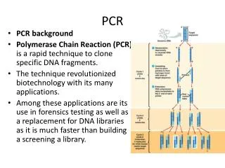
Introduction • With the advent of improved methods for extracting better quality DNA from both paraffin embedded tissues (slides and curls) and fresh tumour tissue, the use of PCR for amplification of specific markers of disease has become a useful diagnostic tool in heamatopathology.
DNA Extraction • In BGL we use a formalin fixed paraffin embedded tissue kit or tumour DNA extraction kit (Qiagen) for histopathology specimens and a routine DNA extraction on our high throughput robot for peripheral blood. • Sample requirements: • 8 10um thick sections or 4 20um thick sections cut on a clean microtome blade and send in a sterile Eppendorf tube. • Fresh snap frozen tumour sample on dry ice • One H+E slide marked clearly with the area of interest plus 10 serial unstained sectionson slides • Peripheral blood or bone marrow (5-10 mls in EDTA)
DNA Extraction Cont….. • Most molecular oncology PCR tests require between 20-100ng DNA dependent on the test eg a simple JAK2V617F test for diagnosis of myeloproliferative neoplasms requires 30ng of DNA in total while a complex multiplex PCR for clonality in lymphoma requires 100ng/ul per multiplex (14 in total) in duplicate which can mean over 1mg for the whole test to be completed!!!! • If we don’t receive the correct amount of sample we may not be able to perform all of the tests required and the quality of the results may be affected by inadequate quantity/quality DNA.
Quality Checking • The quality and quantity of a DNA sample is identified by assessing the 230/260 and 260/280 ratio using the nanadrop (spectrophotometry) Good Quality DNA Poorer Quality DNA
Specimen control ladder • It is also important that we run a specimen control ladder with PET and tumour samples to ensure that the DNA extracted is not too fragmented as this can give false negative results.
Introduction • The majority of lymphoid malignancies belong to the B cell lineage (90-95%) with only 5-7% being T cells • According to van Dongen et al., (2003) the vast majority of lymphoid malignancies (>98%) contain identically (clonal) rearranged immunoglobulin (Ig) and/or T cell receptor (TCR) genes. • Ig and TCR gene loci contain many different variable (V), diversity (D) and joining (J) regions. In Ig heavy chains (IGH), TCRB and TCRD there is an initial D-J rearrangement followed by a V to D-J rearrangement whereas there is only direct V-J rearrangements in IGK, IGL, TCRA and TCRG genes.
Introduction cont…. • During the rearrangements of V, D and J gene segments random deletions and insertions occur at the junction sites. • This results in highly diverse junctional regions, which in turn, leads to an Ig and TCR repertoire of ~10(12).
V genes (124) D genes (27) J genes (6) Constant region VH1 VH2 VH4 VHn Cm Cd Cg Ca JH1 - 6 VH3 VH5 DH1 DH2 DH3 DHn Germline IgH locus Partial DH-JH rearrangement Full VH-DH-JH rearrangement N V N D J CDR3 Random insertion and deletion of nucleotides IGH gene rearrangement
Introduction cont…. • In mature B cells, somatic hypermutation of the V(D)J exon of IgH and Ig light chain genes occurs. • This causes single nucleotide mutations or insertions/deletions to occur. • As such, mature B cell malignancies can show a mutated or unmutated gene profile.
Important points to remember when testing for clonality • Clonality does not always imply malignancy – all results must be interpreted in the context of all of the other available diagnostic criteria. • Ig and TCR gene rearrangements are not markers for lineage – Ig and TCR gene rearrangements are not necessarily restricted to B and T cell lineages, respectively. Cross lineage can occur ie a B cell malignancy can be positive for a TCR gene rearrangement and vice versa. • Limited sensitivity compared to polyclonal background – clonality can only be reported if the clonal peak is 3x higher than the 3rd highest peak in the polyclonal background
BGL and Clonality Testing using IdentiClone gene clonality assay kits from InVivoScribe Technologies DNA sample Liu et al., (2007) DNA size ladder PCR if paraffin-embedded tissue (If DNA > 300 bp) B-cell proliferation T-cell proliferation IGHB + IGKA+B 91% (58%) Initial Screen TCRGA+B 94% (30%) If not clonal TCRγδ+ T-cell lineage unknown TCRαβ+ IGHA+C+D 99% (79%) Extended Screen TCRBA+B 98% (73%) TCRBA+B+C TCRBC + TCRD If not clonal IGL + IGHE 100% (80%) TCRBC + TCRD 100% (82%) If not clonal No evidence of presence of clonal B/T-cell population in the sample by PCR
Patient A Clinical • Sample type: Paraffin embedded tissue • Referred with extensive mediastinal and abdominal lymphadenopathy. Diagnosis hypercalcaemia ?lymphoma • Immunology: A small population of large B cells with the phenotype CD19, CD20 and CD79+, strong Kappa+, CD10, 5, 103- • Cell Pathology Summary: Histology suggestive of diffuse large B cell lymphoma although check immunophenotype and genetic analysis by PCR requested for confirmation. • Immunohistochemistry: Tumour cells +ve with CD20 and BCL2. +ve nuclear staining with BCL6, MUM1,PAX5 and p53
Patient A Clonality Testing IGH Tube B VH(1-7) FR2-JH Clonal peak identified at 271bp +ve Control Polyclonal control Final Diagnosis: Diffuse Large B Cell Lymphoma
Patient B Clinical • Sample Type: Tumour • Referred with ? Lymphoma. Lymphadenopathy left neck • Very difficult lymph node to interpret histologically • Immunohistochemistry: CD10, Cyclin D1 and bcl2 negative • Working diagnosis: Marginal zone lymphoma with follicular colonisation.
Patient B Clonality Testing IGH Tube B VH(1-7) FR2-JH 3rd largest peak in polyclonal background ~1000, clonal peak ~4000 therefore clonal peak over 3x the height of the polyclonal background and reported as weakly clonal in a polyclonal background +ve Control Polyclonal Control Final Diagnosis: Nodal marginal zone lymphoma
Patient C Clinical • Sample: PET • Rt groin biopsy ? Lymphoma – anaemic axillary, inguinal and para-aortic lymphodenopathy – Urgent • Initial histology: reactive hyperplasia but sent to lymphoma expert for opinion who then sent a PET section to BGL for B and T cell clonality testing • Concurrently, a peripheral blood sample was sent for JAK2 V617F testing – no mutation present. • Immunology showed a 1-2% population of Lambda +ve cells • Initial B and T cell screen on PET = no clonality identified = reactive rather than malignant • Further testing, an initial B and T cell screen on peripheral blood showed……….
Patient C Clonality Testing (PET and Peripheral blood) TCRG (tube A) VγIf Vγ10 – Jγ1.3/2.3 + Jγ1.1/2.1 Negative for the detection of clonal TCRG chain rearrangements PET 3rd largest peak in polyclonal background ~25000, clonal peak ~75000 therefore clonal peak over 3x the height of the polyclonal background and reported as weakly clonal in a polyclonal background PB +VE PC
What went wrong with the clonality testing? • Nothing!! • We reported that in the context of overall diagnostic criteria, clonal cell populations can indicate the presence of haematological malignancy. • While monoclonality is a key feature of tumour cell populations it does not always imply malignancy because some reactive processes contain large clonal lymphocyte populations. Therefore we need to take into consideration the whole clinical picture rather than just the result of the clonality testing. • The patients flow results showed a 1-2% population of malignant cells which were Lambda +ve, this is part of the extended panel which also includes other B cell markers (Framework 1 and 3 and the incomplete DH’s) • However, its absence would not detract from the clonality already identified in the T cells
Patient C Cont…. • An extended B cell screen was initiated and was negative for the detection of IGH rearrangements • A second core biopsy was taken. The histology showed HHV8 (human herpes virus-8) positive Kaposi’s sarcoma and HHV8 associated lymphoproliferative disorder. HIV negative patient, probably common variable immunodeficiency. • HHV8 positive plasma cells are monotypic but polyclonal. (On review first biopsy also HHV8+ but not Castlemans Disease)) • Patient now treated with Rituximab (anti-CD20 monoclonal antibody therapy) and responding well
Allele Specific PCR • Amplification of specific alleles, or DNA sequence variants, at the same locus. Specificity is achieved by designing one or both PCR primers so that they partially overlap the site of sequence difference between the amplified alleles. • Used in haemato-oncology for detection of JAK2V617F mutation in myleoproliferative neoplasms. • GGA GTA TGT GTC T (WT) • GGA GTA TGT TTC T (Mut) Pos = White Neg = Yellow
Capillary Electrophoresis • Gel-capillary electrophoresis is an alternative to traditional electrophoretic gel based techniques. • Many advantages including excellent resolution which enables multiplex reactions (ie more than one set of primers in each reaction mix) reduced sample quantities, and automation. • Used as the detection method for many molecular oncology tests including clonality testing in lymphoma and Loss of heterozygosity in oligodendrogliomas Blood Tumour
Logarithmic dilution series Day 28 samples Positive Control test 10(6) 10(5) 10(3) 10(2) 10(1) Real-time quantitative PCR (RQ-PCR) • Quantitative real-time polymerase chain reaction (PCR) provides an accurate method for determination of levels of specific DNA and RNA (RT) sequences in tissue samples. It is based on detection of a fluorescent signal produced proportionally during amplification of a PCR product. • Usedfor quantification of BCR-ABL molecules in chronic myeloid leukaemia (left) and minimal residual disease detection in acute lymphoblastic leukaemia (right)
Sequencing • DNA sequencing is the process of determining the nucleotide order of a given DNA fragment. • Most automated laboratories use in dye-terminator sequencing, where each of the four dideoxynucleotide chain terminators (A,C,G,T) is labelled with a fluorescent dye with a different wavelength and is compared to a wild type sequence for identification of changes in the sequence. 4bp inserted duplication in the NPM1 gene in AML
Pyrosequencing • High throughput method of DNA sequencing based on a “sequence by synthesis” principle. In short, this method synthesizes the complementary strand of a single strand of DNA one base pair at a time. • When the complementary base binds to the single stranded DNA a chemiluminescent signal is given off which can be detected, allowing the determination of the sequence in real time. • In BGL this technology will be developed to allow for screening of mutations associated with myeloproliferative neoplasms (for example JAK2V617F and MPLW515K/L), K-Ras testing in colorectal carcinoma and methylation status in glioblastoma brain tumours.
High resolution melt analysis • Perform a PCR using primers specific for gene in question • Following the PCR, heat the amplicon DNA from around 50˚C up to around 95˚C. • At some point during this process, the melting temperature of the amplicon is reached and the two strands of DNA separate or “melt” apart • The dsDNA is labelled with a fluorescent dye, which fluoresces brightly. As the stands separate the intensity of the dye is reduced and this reduction is measured in real time producing a melt curve
High resolution melt analysis cont.. • BGL is currently developing this technology for testing of MPL and JAK2 Exon 12 mutations in MPNs but many papers have used it for BRCA1 and BRCA2 mutation screening and TP53 screening in breast and ovarian cancer which we are ?hoping to develop in the future. http://en.wikipedia.org/wiki/High_Resolution_Melt
References • Van Dongen et al., Design and standardisation of PCR primers and protocols for detection of clonal immunoglobulin and T cell receptor gene recombinations in suspect lymphoproliferations. Leukemia, 2003. 17: 2257-2317 • Liu H et al., A practical study for the routine use of BIOMED-2 PCR assays for the detection of B and T cell clonality in diagnostic haematopathology. Br J Haematol. 2007. 138(1):31-43
Acknowledgements • Eileen Roberts (Head of Department, BGL) • Dr Jerry Hancock (Molecular Oncology, BGL) • Helen, Rich, Paul, Jess and Adelea (Molecular Oncology team) • Dr Nicholas Rooney (Cellular Pathology) • Konstantin Sidelnikov (InVivoScribe Technologies, Inc)